
KNOWPIA
WELCOME TO KNOWPIA

Anterior nasal spine 
Summary
The anterior nasal spine, or anterior nasal spine of maxilla, is a bony projection in the skull that serves as a cephalometric landmark.[1] The anterior nasal spine is the projection formed by the fusion of the two maxillary bones at the intermaxillary suture. It is placed at the level of the nostrils, at the uppermost part of the philtrum. It rarely fractures.[2]
| Anterior nasal spine | |
|---|---|
 Left maxilla. Nasal surface (anterior nasal spine labeled at bottom right) | |
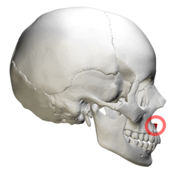 The skull from the side. Anterior nasal spine is at right (shown in red). | |
| Details | |
| Identifiers | |
| Latin | spina nasalis anterior maxillae |
| TA98 | A02.1.12.011 |
| TA2 | 766 |
| FMA | 75770 |
| Anatomical terms of bone [edit on Wikidata] | |
Additional images edit
-
 Animation. Anterior nasal spine shown in red.
 -
 Left maxilla. Anterior nasal spine shown in red.
 -
 Skull. Anterior view. Anterior nasal spine shown in red.
 -
 Right maxilla. Anterior nasal spine labeled at center left.


See also edit
References edit
![]() This article incorporates text in the public domain from page 158 of the 20th edition of Gray's Anatomy (1918)
This article incorporates text in the public domain from page 158 of the 20th edition of Gray's Anatomy (1918)
- ^ Albarakati, SF; Kula, KS; Ghoneima, AA (January 2012). "The reliability and reproducibility of cephalometric measurements: a comparison of conventional and digital methods". Dento Maxillo Facial Radiology. 41 (1): 11–7. doi:10.1259/dmfr/37010910. PMC 3520271. PMID 22184624.
- ^ Knipe, Henry. "Anterior nasal spine fracture | Radiology Case | Radiopaedia.org". radiopaedia.org. Retrieved 13 October 2018.
External links edit
Wikimedia Commons has media related to Anterior nasal spine.
- Diagram at upstate.edu - side
- Diagram at upstate.edu - front
- Anatomy photo:22:os-1911 at the SUNY Downstate Medical Center - "Osteology of the Skull: The Maxilla"
- "Anatomy diagram: 34256.000-1". Roche Lexicon - illustrated navigator. Elsevier. Archived from the original on 2012-12-27.
- "Anatomy diagram: 34256.000-2". Roche Lexicon - illustrated navigator. Elsevier. Archived from the original on 2013-06-11.