


Summary
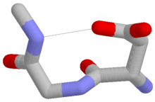
The Asx turn[1][2][3][4][5][6][7] is a structural feature in proteins and polypeptides. It consists of three amino acid residues (labeled i, i+1 and i+2) in which residue i is an aspartate (Asp) or asparagine (Asn) that forms a hydrogen bond from its sidechain CO group to the mainchain NH group of residue i+2. About 14% of Asx residues present in proteins belong to Asx turns.
The name "Asx" is used here to represent either of the amino acids aspartate (Asp) or asparagine (Asn).
Types edit
Four types of Asx turn can be distinguished:[8] types I, I’, II and II’. These categories correspond to those of the better-known hydrogen-bonded beta turns, which have four residues and a hydrogen bond between the CO of residue i and the NH of residue i+3. Asx turns and beta turns have structurally similar hydrogen-bonded loops and exhibit sidechain-mainchain mimicry in the sense that the sidechain of residue i of the Asx turn mimics the mainchain of residue i of the beta turn. Regarding their occurrence in proteins, they differ in that type I is the commonest of the four beta turns while type II’ is the commonest of the Asx turns.
Occurrence edit
Asx and ST turns both occur frequently at the N-termini of α-helices.[9][10][11][12] as part of Asx motifs or ST motifs such that the Asx, serine or threonine is the N cap residue. They are thus often regarded as helix capping features.
Related motifs edit
Similar motifs occur with serine or threonine as residue i, which are called ST turns.[13] In spite of serine and threonine having one less sidechain atom, such that the sidechain-mainchain mimicry of β turns is imperfect, these features occur in proteins as the four types in numbers approaching those of Asx turns. They also exhibit a tendency to substitute each other over evolutionary time.[14]
A proportion of Asx turns are accompanied by a mainchain–mainchain hydrogen bond that qualifies them as Asx motifs.
References edit
- ^ Richardson, JS (1981). "The anatomy and taxonomy of protein structure". Advances in Protein Chemistry Volume 34. Vol. 34. pp. 167–339. doi:10.1016/S0065-3233(08)60520-3. ISBN 9780120342341. PMID 7020376.
- ^ Tainer, JA; Getzoff ED (1982). "Determination and analysis of the 2 A-structure of copper, zinc superoxide dismutase". Journal of Molecular Biology. 160 (2): 181–217. doi:10.1016/0022-2836(82)90174-7. PMID 7175933.
- ^ Rees, DC; Lewis M (1983). "Refined crystal structure of carboxypeptidase a at 1.54 Å resolution". Journal of Molecular Biology. 168 (2): 367–387. doi:10.1016/S0022-2836(83)80024-2. PMID 6887246.
- ^ Eswar, N; Ramachandran C (1999). "Secondary structures without backbone: An analysis of backbone mimicry by polar side chains in proteins". Protein Engineering. 12 (6): 447–455. doi:10.1093/protein/12.6.447. PMID 10388841.
- ^ Chakrabarti, P; Pal D (2001). "Interrelationships of side-chain and main-chain conformations in proteins". Progress in Biophysics and Molecular Biology. 76 (1–2): 1–102. doi:10.1016/s0079-6107(01)00005-0. PMID 11389934.
- ^ Duddy, WJ; Nissink WMJ; Allen, Frank H.; Milner-White, E. James (2004). "Mimicry by asx- and ST-turns of the four main types of β-turn in proteins". Protein Science. 13 (11): 3051–3055. doi:10.1110/ps.04920904. PMC 2286581. PMID 15459339.
- ^ Thakur, AK; Kishore R (2006). "Characterization of β-turn and asx-turns mimicry in a model peptide : Stabilization via C-H•••O interaction". Biopolymers. 81 (6): 440–449. doi:10.1002/bip.20441. PMID 16411188. S2CID 27091571.
- ^ Duddy, WJ; Nissink WMJ; Allen, Frank H.; Milner-White, E. James (2004). "Mimicry by asx- and ST-turns of the four main types of beta turn in proteins". Protein Science. 13 (11): 3051–3055. doi:10.1110/ps.04920904. PMC 2286581. PMID 15459339.
- ^ Doig, AJ; Macarthur MW; MacArthur, Malcolm W.; Thornton, Janet M. (1997). "Structures of N-termini of helices in proteins". Protein Science. 6 (1): 147–155. doi:10.1002/pro.5560060117. PMC 2143508. PMID 9007987.
- ^ Presta, LG; Rose GD (1988). "Helix Caps". Science. 240 (4859): 1632–1641. Bibcode:1988Sci...240.1632P. doi:10.1126/science.2837824. PMID 2837824.
- ^ Aurora, R; Rose GD (1998). "Helix Capping". Protein Science. 7 (1): 21–38. doi:10.1002/pro.5560070103. PMC 2143812. PMID 9514257.
- ^ Gunasekaran, K; Nagarajam HA; Ramakrishnan, C; Balaram, P (1998). "Stereochemical punctuation marks in protein structure". Journal of Molecular Biology. 275 (5): 917–932. doi:10.1006/jmbi.1997.1505. PMID 9480777. S2CID 35919397.
- ^ Duddy, WJ; Nissink WMJ; Allen, Frank H.; Milner-White, E. James (2004). "Mimicry by asx- and ST-turns of the four main types of β-turn in proteins". Protein Science. 13 (11): 3051–3055. doi:10.1110/ps.04920904. PMC 2286581. PMID 15459339.
- ^ Wan, W-Y; Milner-White EJ (2009). "A Recurring Two-Hydrogen-bond Motif Incorporating a Serine or Threonine Residue is found both at α-Helical N Termini and in Other Situations". Journal of Molecular Biology. 286 (5): 1651–1662. doi:10.1006/jmbi.1999.2551. PMID 10064721.
External links edit
- ^ Leader, DP; Milner-White EJ (2009). "Motivated Proteins: A web application for studying small three-dimensional protein motifs". BMC Bioinformatics. 10: 60. doi:10.1186/1471-2105-10-60. PMC 2651126. PMID 19210785.
- ^ Golovin, A; Henrick K (2008). "MSDmotif: exploring protein sites and motifs". BMC Bioinformatics. 9: 312. doi:10.1186/1471-2105-9-312. PMC 2491636. PMID 18637174.