


Summary
The basilar part of the occipital bone (also basioccipital) extends forward and upward from the foramen magnum, and presents in front an area more or less quadrilateral in outline.
| Basilar part of occipital bone | |
|---|---|
 Occipital bone inner surface (basilar part is shown in red) | |
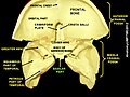
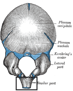 Occipital bone outer surface, at birth (basilar part is at bottom) | |
| Details | |
| Identifiers | |
| Latin | pars basilaris ossis occipitalis |
| TA98 | A02.1.04.005 |
| TA2 | 554 |
| FMA | 52858 |
| Anatomical terms of bone [edit on Wikidata] | |
In the young skull, this area is rough and uneven, and is joined to the body of the sphenoid by a plate of cartilage.
By the twenty-fifth year, this cartilaginous plate is ossified, and the occipital and sphenoid form a continuous bone.
Surfaces edit
On its lower surface, about 1 cm. in front of the foramen magnum, is the pharyngeal tubercle which gives attachment to the fibrous raphe of the pharynx.
On either side of the middle line the longus capitis and rectus capitis anterior are inserted, and immediately in front of the foramen magnum the anterior atlantooccipital membrane is attached.
The upper surface, which constitutes the lower half of the clivus, presents a broad, shallow groove which inclines upward and forward from the foramen magnum; it supports the medulla oblongata, and near the margin of the foramen magnum gives attachment to the tectorial membrane
On the lateral margins of this surface are faint grooves for the inferior petrosal sinuses.
Additional images edit
-
 Occipital bone. Basilar part shown in red.
 -
 Human skull seen from below. Basilar part shown in red.
 -
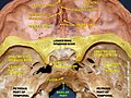
 Human skull seen from above (parietal bones have been removed). Basilar part shown in red.
 -
 Occipital bone. Outer surface.
 -
 Membrana tectoria, transverse, and alar ligaments.
 -
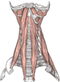 The anterior vertebral muscles.
 -
 Basilar part of occipital bone
 -
 Basilar part of occipital bone
 -
 Basilar part of occipital bone
 -
 Tympanic cavity. Facial canal. Internal carotid artery.


References edit
![]() This article incorporates text in the public domain from page 132 of the 20th edition of Gray's Anatomy (1918)
This article incorporates text in the public domain from page 132 of the 20th edition of Gray's Anatomy (1918)
External links edit
- Anatomy photo:22:os-0913 at the SUNY Downstate Medical Center