


Summary
Bifidobacterium is a genus of gram-positive, nonmotile, often branched anaerobic bacteria. They are ubiquitous inhabitants of the gastrointestinal tract[2][3] though strains have been isolated from the vagina[4] and mouth (B. dentium) of mammals, including humans. Bifidobacteria are one of the major genera of bacteria that make up the gastrointestinal tract microbiota in mammals. Some bifidobacteria are used as probiotics.
| Bifidobacterium | |
|---|---|

| |
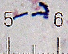
| Bifidobacterium adolescentis | |
| Scientific classification | |
| Domain: | Bacteria |
| Phylum: | Actinomycetota |
| Class: | Actinomycetia |
| Order: | Bifidobacteriales |
| Family: | Bifidobacteriaceae |
| Genus: | BifidobacteriumOrla-Jensen 1924 (Approved Lists 1980)[1] |
| Type species | |
| Bifidobacterium bifidum (Tissier 1900) Orla-Jensen 1924 (Approved Lists 1980)
| |
| Species | |
|
See text. | |
Before the 1960s, Bifidobacterium species were collectively referred to as Lactobacillus bifidus.
History edit
In 1899, Henri Tissier, a French pediatrician at the Pasteur Institute in Paris, isolated a bacterium characterised by a Y-shaped morphology ("bifid") in the intestinal microbiota of breast-fed infants and named it "bifidus".[5] In 1907, Élie Metchnikoff, deputy director at the Pasteur Institute, propounded the theory that lactic acid bacteria are beneficial to human health.[5] Metchnikoff observed that the longevity of Bulgarians was the result of their consumption of fermented milk products.[6] Metchnikoff also suggested that "oral administration of cultures of fermentative bacteria would implant the beneficial bacteria in the intestinal tract".[7]
Metabolism edit
The genus Bifidobacterium possesses a unique fructose-6-phosphate phosphoketolase pathway employed to ferment carbohydrates.[citation needed]
Much metabolic research on bifidobacteria has focused on oligosaccharide metabolism, as these carbohydrates are available in their otherwise nutrient-limited habitats. Infant-associated bifidobacterial phylotypes appear to have evolved the ability to ferment milk oligosaccharides, whereas adult-associated species use plant oligosaccharides, consistent with what they encounter in their respective environments. As breast-fed infants often harbor bifidobacteria-dominated gut consortia, numerous applications attempt to mimic the bifidogenic properties of milk oligosaccharides. These are broadly classified as plant-derived fructooligosaccharides or dairy-derived galactooligosaccharides, which are differentially metabolized and distinct from milk oligosaccharide catabolism.[3]
Response to oxygen edit
The sensitivity of members of the genus Bifidobacterium to O2 generally limits probiotic activity to anaerobic habitats. Recent research has reported that some Bifidobacterium strains exhibit various types of oxic growth. Low concentrations of O2 and CO2 can have a stimulatory effect on the growth of these Bifidobacterium strains. Based on the growth profiles under different O2 concentrations, the Bifidobacterium species were classified into four classes: O2-hypersensitive, O2-sensitive, O2-tolerant, and microaerophilic. The primary factor responsible for aerobic growth inhibition is proposed to be the production of hydrogen peroxide (H2O2) in the growth medium. A H2O2-forming NADH oxidase was purified from O2-sensitive Bifidobacterium bifidum and was identified as a b-type dihydroorotate dehydrogenase. The kinetic parameters suggested that the enzyme could be involved in H2O2 production in highly aerated environments.[8]
Genomes edit
Members of the genus Bifidobacterium have genome sizes ranging from 1.73 (Bifidobacterium indicum) to 3.25 Mb (Bifidobacterium biavatii), corresponding to 1,352 and 2,557 predicted protein-encoding open reading frames, respectively.[9]
Functional classification of Bifidobacterium genes, including the pan-genome of this genus, revealed that 13.7% of the identified bifidobacterial genes encode enzymes involved in carbohydrate metabolism.[9]
Clinical uses edit
Adding Bifidobacterium as a probiotic to conventional treatment of ulcerative colitis has been shown to be associated with improved rates of remission and improved maintenance of remission.[10] Some Bifidobacterium strains are considered as important probiotics and used in the food industry. Different species and/or strains of bifidobacteria may exert a range of beneficial health effects, including the regulation of intestinal microbial homeostasis, the inhibition of pathogens and harmful bacteria that colonize and/or infect the gut mucosa, the modulation of local and systemic immune responses, the repression of procarcinogenic enzymatic activities within the microbiota, the production of vitamins, and the bioconversion of a number of dietary compounds into bioactive molecules.[3] Bifidobacteria improve the gut mucosal barrier and lower levels of lipopolysaccharide in the intestine.[11]
Bifidobacteria may also improve abdominal pain in patients with irritable bowel syndrome (IBS) though studies to date have been inconclusive.[12]
Naturally occurring Bifidobacterium spp. may discourage the growth of Gram-negative pathogens in infants.[13]
Mother's milk contains high concentrations of lactose and lower quantities of phosphate (pH buffer). Therefore, when mother's milk is fermented by lactic acid bacteria (including bifidobacteria) in the infant's gastrointestinal tract, the pH may be reduced, making it more difficult for Gram-negative bacteria to grow.[citation needed]
Bifidobacteria and the infant gut edit
The human infant gut is relatively sterile up until birth, where it takes up bacteria from its surrounding environment and its mother.[14] The microbiota that makes up the infant gut differs from the adult gut. An infant reaches the adult stage of their microbiome at around three years of age, when their microbiome diversity increases, stabilizes, and the infant switches over to solid foods. Breast-fed infants are colonized earlier by Bifidobacterium when compared to babies that are primarily formula-fed.[15] Bifidobacterium is the most common bacteria in the infant gut microbiome.[16] There is more variability in genotypes over time in infants, making them less stable compared to the adult Bifidobacterium. Infants and children under three years old show low diversity in microbiome bacteria, but more diversity between individuals when compared to adults.[17] Reduction of Bifidobacterium and increase in diversity of the infant gut microbiome occurs with less breast-milk intake and increase of solid food intake. Mammalian milk all contain oligosaccharides showing natural selection [clarification needed]. Human milk oligosaccharides are not digested by enzymes and remain whole through the digestive tract before being broken down in the colon by microbiota. Bifidobacterium species genomes of B. longum, B. bifidum, B. breve contain genes that can hydrolyze some of the human milk oligosaccharides and these are found in higher numbers in infants that are breast-fed. Glycans that are produced by the humans are converted into food and energy for the B. bifidum. showing an example of coevolution.[18]
Species edit
The genus Bifidobacterium comprises the following species:[19]
- B. actinocoloniiforme Killer et al. 2011
- B. adolescentis Reuter 1963 (Approved Lists 1980)
- B. aemilianum Alberoni et al. 2019
- B. aerophilum Michelini et al. 2017
- B. aesculapii Modesto et al. 2014
- B. amazonense Lugli et al. 2021
- B. angulatum Scardovi and Crociani 1974 (Approved Lists 1980)
- B. animalis (Mitsuoka 1969) Scardovi and Trovatelli 1974 (Approved Lists 1980)
- B. anseris Lugli et al. 2018
- B. apousia Chen et al. 2022
- B. apri Pechar et al. 2017
- B. aquikefiri Laureys et al. 2016
- B. asteroides Scardovi and Trovatelli 1969 (Approved Lists 1980)
- B. avesanii Michelini et al. 2019
- B. biavatii Endo et al. 2012
- B. bifidum (Tissier 1900) Orla-Jensen 1924 (Approved Lists 1980)
- B. bohemicum Killer et al. 2011
- B. bombi Killer et al. 2009
- B. boum Scardovi et al. 1979 (Approved Lists 1980)
- B. breve Reuter 1963 (Approved Lists 1980)
- B. callimiconis Duranti et al. 2019
- B. callitrichidarum Modesto et al. 2018
- B. callitrichos Endo et al. 2012
- B. canis Neuzil-Bunesova et al. 2020
- B. castoris Duranti et al. 2019
- B. catenulatum Scardovi and Crociani 1974 (Approved Lists 1980)
- B. catulorum Modesto et al. 2018
- B. cebidarum Duranti et al. 2020
- B. choerinum Scardovi et al. 1979 (Approved Lists 1980)
- B.choladohabitans Chen et al. 2022
- B. choloepi Modesto et al. 2020
- B. colobi Lugli et al. 2021
- B. commune Praet et al. 2015
- B. criceti Lugli et al. 2018
- "B. crudilactis" Delcenserie et al. 2007
- B.cuniculi Scardovi et al. 1979 (Approved Lists 1980)
- B. dentium Scardovi and Crociani 1974 (Approved Lists 1980)
- B. dolichotidis Duranti et al. 2019
- "B. eriksonii" Cato et al. 1970
- B. erythrocebi Neuzil-Bunesova et al. 2021
- B. eulemuris Michelini et al. 2016
- B. faecale Choi et al. 2014
- B. felsineum Modesto et al. 2020
- B. gallicum Lauer 1990
- B. gallinarum Watabe et al. 1983
- B. globosum (ex Scardovi et al. 1969) Biavati et al. 1982
- B. goeldii Duranti et al. 2019
- B. hapali Michelini et al. 2016
- B. Lugli et al. 2018
- B. indicum Scardovi and Trovatelli 1969 (Approved Lists 1980)
- B. italicum Lugli et al. 2018
- B. jacchi Modesto et al. 2019
- B. lemurum Modesto et al. 2015
- B. leontopitheci Duranti et al. 2020
- B. longum Reuter 1963 (Approved Lists 1980)
- B. magnum Scardovi and Zani 1974 (Approved Lists 1980)
- B.margollesii Lugli et al. 2018
- B. merycicum Biavati and Mattarelli 1991
- B. miconis Lugli et al. 2021
- B. miconisargentati Lugli et al. 2021
- B. minimum Biavati et al. 1982
- B. mongoliense Watanabe et al. 2009
- B. moraviense Neuzil-Bunesova et al. 2021
- B. moukalabense Tsuchida et al. 2014
- B. myosotis Michelini et al. 2016
- B. oedipodis Neuzil-Bunesova et al. 2021
- B. olomucense Neuzil-Bunesova et al. 2021
- B. panos Neuzil-Bunesova et al. 2021
- B. parmae Lugli et al. 2018
- "B. platyrrhinorum" Modesto et al. 2020
- B. pluvialisilvae Lugli et al. 2021
- B. polysaccharolyticum Chen et al. 2022
- B. pongonis Lugli et al. 2021
- B. porcinum (Zhu et al. 2003) Nouioui et al. 2018
- B. primatium Modesto et al. 2020
- B. pseudocatenulatum Scardovi et al. 1979 (Approved Lists 1980)
- B. pseudolongum Mitsuoka 1969 (Approved Lists 1980)
- B. psychraerophilum Simpson et al. 2004
- B. pullorum Trovatelli et al. 1974 (Approved Lists 1980)
- B. ramosum Michelini et al. 2017
- B. reuteri Endo et al. 2012
- B. rousetti Modesto et al. 2021
- "B. ruminale" Scardovi et al. 1969
- B. ruminantium Biavati and Mattarelli 1991
- B. saguini Endo et al. 2012
- B. saguinibicoloris Lugli et al. 2021
- "B. saimiriisciurei" Modesto et al. 2020
- B. samirii Duranti et al. 2019
- B. santillanense Lugli et al. 2021
- B. scaligerum Modesto et al. 2020
- B. scardovii Hoyles et al. 2002
- B. simiarum Modesto et al. 2020
- B. simiiventris Lugli et al. 2021
- B. stellenboschense Endo et al. 2012
- B. subtile Biavati et al. 1982
- B. thermacidophilum Dong et al. 2000
- B. thermophilum corrig. Mitsuoka 1969 (Approved Lists 1980)
- B. tibiigranuli Eckel et al. 2020
- B. tissieri corrig. Michelini et al. 2016
- B. tsurumiense Okamoto et al. 2008
- "B. urinalis" Hojo et al. 2007
- B. vansinderenii Duranti et al. 2017
- B. vespertilionis Modesto et al. 2021
- B. xylocopae Alberoni et al. 2019
See also edit
References edit
- ^ Orla-Jensen S. (1924). "Classification des bactéries lactiques" [Classification of the lactic acid bacteria]. Le Lait. 4 (36): 468–474. doi:10.1051/lait:19243627.
- ^ Schell MA, Karmirantzou M, Snel B, Vilanova D, Berger B, Pessi G, Zwahlen MC, Desiere F, Bork P, Delley M, Pridmore RD, Arigoni F (October 2002). "The genome sequence of Bifidobacterium longum reflects its adaptation to the human gastrointestinal tract". Proceedings of the National Academy of Sciences of the United States of America. 99 (22): 14422–7. Bibcode:2002PNAS...9914422S. doi:10.1073/pnas.212527599. PMC 137899. PMID 12381787.
- ^ a b c Mayo B, van Sinderen D, eds. (2010). Bifidobacteria: Genomics and Molecular Aspects. Caister Academic Press. ISBN 978-1-904455-68-4.[page needed]
- ^ Albert, Arianne Y. K.; Chaban, Bonnie; Wagner, Emily C.; Schellenberg, John J.; Links, Matthew G.; Schalkwyk, Julie van; Reid, Gregor; Hemmingsen, Sean M.; Hill, Janet E.; Money, Deborah; Group, VOGUE Research (12 August 2015). "A Study of the Vaginal Microbiome in Healthy Canadian Women Utilizing cpn60-Based Molecular Profiling Reveals Distinct Gardnerella Subgroup Community State Types". PLOS ONE. 10 (8): e0135620. Bibcode:2015PLoSO..1035620A. doi:10.1371/journal.pone.0135620. PMC 4534464. PMID 26266808.
- ^ a b "Potential of probiotics as biotherapeutic agents targeting the innate immune system" (PDF). African Journal of Biotechnology. February 2005.
- ^ "Probiotics: 100 years (1907–2007) after Elie Metchnikoff's Observation" (PDF). Communicating Current Research and Educational Topics and Trends in Applied Microbiology. February 2007. Archived from the original (PDF) on 2012-10-04.
- ^ "Pioneers of Probiotics". European Probiotic Association. February 2012. Archived from the original on 2013-07-22. Retrieved 2013-07-01.
- ^ Sonomoto K, Yokota A, eds. (2011). Lactic Acid Bacteria and Bifidobacteria: Current Progress in Advanced Research. Caister Academic Press. ISBN 978-1-904455-82-0.[page needed]
- ^ a b Milani C, Turroni F, Duranti S, Lugli GA, Mancabelli L, Ferrario C, van Sinderen D, Ventura M (February 2016). "Genomics of the Genus Bifidobacterium Reveals Species-Specific Adaptation to the Glycan-Rich Gut Environment". Applied and Environmental Microbiology. 82 (4): 980–991. Bibcode:2016ApEnM..82..980M. doi:10.1128/AEM.03500-15. PMC 4751850. PMID 26590291.
- ^ Ghouri YA, Richards DM, Rahimi EF, Krill JT, Jelinek KA, DuPont AW (9 December 2014). "Systematic review of randomized controlled trials of probiotics, prebiotics, and synbiotics in inflammatory bowel disease". Clinical and Experimental Gastroenterology. 7: 473–87. doi:10.2147/CEG.S27530. PMC 4266241. PMID 25525379.
- ^ Pinzone MR, Celesia BM, Di Rosa M, Cacopardo B, Nunnari G (2012). "Microbial translocation in chronic liver diseases". International Journal of Microbiology. 2012: 694629. doi:10.1155/2012/694629. PMC 3405644. PMID 22848224.
- ^ Pratt, Charlotte; Campbell, Matthew D. (2019-11-18). "The Effect of Bifidobacterium on Reducing Symptomatic Abdominal Pain in Patients with Irritable Bowel Syndrome: A Systematic Review". Probiotics and Antimicrobial Proteins. 12 (3): 834–839. doi:10.1007/s12602-019-09609-7. ISSN 1867-1306. PMC 7456408. PMID 31741311.
- ^ Liévin V, Peiffer I, Hudault S, Rochat F, Brassart D, Neeser JR, Servin AL (November 2000). "Bifidobacterium strains from resident infant human gastrointestinal microflora exert antimicrobial activity". Gut. 47 (5): 646–52. doi:10.1136/gut.47.5.646. PMC 1728100. PMID 11034580.
- ^ Pham VT, Lacroix C, Braegger CP, Chassard C (July 2016). "Early colonization of functional groups of microbes in the infant gut". Environmental Microbiology. 18 (7): 2246–58. Bibcode:2016EnvMi..18.2246P. doi:10.1111/1462-2920.13316. PMID 27059115.
- ^ Bourlieu C, Bouzerzour K, FerretBernard S, Bourgot CL, Chever S, Menard O, Deglaire A, Cuinet I, Ruyet PL, Bonhomme C, Dupont D (2015). "Infant formula interface and fat source impact on neonatal digestion and gut microbiota". European Journal of Lipid Science and Technology. 117 (10): 1500–1512. doi:10.1002/ejlt.201500025. ISSN 1438-9312.
- ^ Turroni F, Peano C, Pass DA, Foroni E, Severgnini M, Claesson MJ, Kerr C, Hourihane J, Murray D, Fuligni F, Gueimonde M, Margolles A, De Bellis G, O'Toole PW, van Sinderen D, Marchesi JR, Ventura M (2012-05-11). "Diversity of bifidobacteria within the infant gut microbiota". PLOS ONE. 7 (5): e36957. Bibcode:2012PLoSO...736957T. doi:10.1371/journal.pone.0036957. PMC 3350489. PMID 22606315.
- ^ Matamoros S, Gras-Leguen C, Le Vacon F, Potel G, de La Cochetiere MF (April 2013). "Development of intestinal microbiota in infants and its impact on health". Trends in Microbiology. 21 (4): 167–73. doi:10.1016/j.tim.2012.12.001. PMID 23332725.
- ^ Turroni F, Milani C, Duranti S, Ferrario C, Lugli GA, Mancabelli L, van Sinderen D, Ventura M (January 2018). "Bifidobacteria and the infant gut: an example of co-evolution and natural selection". Cellular and Molecular Life Sciences. 75 (1): 103–118. doi:10.1007/s00018-017-2672-0. PMID 28983638. S2CID 24103287.
- ^ Euzéby JP, Parte AC. "Actinomycetaceae". List of Prokaryotic names with Standing in Nomenclature (LPSN). Retrieved June 17, 2021.
External links edit
- Bifidobacterium at Microbe Wiki
- Genomes Online Database contains many Bifidobacterium genome projects
- Comparative Analysis of Bifidobacterium Genomes (at DOE's IMG system)
- Bifidobacterium at BacDive - the Bacterial Diversity Metadatabase
- Spotlight on Bifidobacteria