


Summary
CD83 (Cluster of Differentiation 83) is a human protein encoded by the CD83 gene.[5]
| CD83 | |||||||||||||||||||||||||||||||||||||||||||||||||||
|---|---|---|---|---|---|---|---|---|---|---|---|---|---|---|---|---|---|---|---|---|---|---|---|---|---|---|---|---|---|---|---|---|---|---|---|---|---|---|---|---|---|---|---|---|---|---|---|---|---|---|---|
| Identifiers | |||||||||||||||||||||||||||||||||||||||||||||||||||
| Aliases | CD83, BL11, HB15, CD83 molecule | ||||||||||||||||||||||||||||||||||||||||||||||||||
| External IDs | OMIM: 604534 MGI: 1328316 HomoloGene: 3121 GeneCards: CD83 | ||||||||||||||||||||||||||||||||||||||||||||||||||
| |||||||||||||||||||||||||||||||||||||||||||||||||||
| |||||||||||||||||||||||||||||||||||||||||||||||||||
| |||||||||||||||||||||||||||||||||||||||||||||||||||
| |||||||||||||||||||||||||||||||||||||||||||||||||||
| |||||||||||||||||||||||||||||||||||||||||||||||||||
| Wikidata | |||||||||||||||||||||||||||||||||||||||||||||||||||
| |||||||||||||||||||||||||||||||||||||||||||||||||||


Structure edit
The membrane-bound form of CD83 consists of an extracellular V-type immunoglobulin-like domain, a transmembrane domain and a cytoplasmic signaling tail. A free soluble form consists of the immunoglobulin-like domain alone. Membrane-bound CD83 is expected to form trimers. Soluble CD83 is able to assemble into dodecameric complexes.[6]
Gene edit
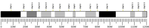
The CD83 gene is located on human chromosome 6p23 and mouse chromosome 13. In humans, a promoter 261 bp upstream consists of five NF-κB and three interferon regulatory factor binding sites, reflecting the involvement of CD83 in inflammation,[7] as well as binding sites for the aryl hydrocarbon receptor. The latter also occur in an enhancer sequence located 185 bp downstream, inside the second intron,[8] and may suggest negative regulation of transcription by microbial metabolites produced in the gut.
Function edit
The transmembrane domain of membrane-bound CD83 stabilizes MHC II, costimulatory molecules and CD28 in the membrane by antagonizing MARCH-family E3 ubiquitin ligases.[9][10]
Ligands edit
It is not clear what ligands interact with CD83, but membrane-bound CD83 may homotypically interact with the soluble form, suggesting autocrine immune regulation.[11] However, it contrasts with differences between the single expression of soluble CD83 on monocytes and membrane-bound CD83 on activated dendritic cells seems also as their good marker.[clarification needed][12] Soluble CD83 also binds to CD154, leading to T helper type 2 lymphocyte apoptosis by suppression of Bcl-2 inhibitors.[13]
Positive selection edit
The development of thymocytes during the positive-selection stage may be guided by CD83 expression on cortical thymic epithelial cells (cTECs). CD4+CD8+ double-positive thymocytes surrounded by specially differentiated cTECs called thymic nurse cells are tested for function of their αβ T cell receptor (TCR); a nonreactive TCR leads to thymocyte death by neglect. Successful rearrangement of a reactive TCR supports survival and restriction of expression to CD4 or CD8 alone on single-positive thymocytes, depending on the ability to recognize MHC II or MHC I, respectively. Upregulation of MHC II turnover on thymic nurse cells by CD83 may enlarge the population of CD4+ single-positive thymocytes.[14][10]
Regulatory T cells edit

T regulatory cells (Treg cells) are present in two major populations: thymically induced and peripherally induced Treg cells. All Treg cells express the Foxp3 transcription factor, establishing their suppressive phenotype. Foxp3 expression is not affected by loss of CD83 in a CD83 knockout mouse. In contrast, CD83 seems important for peripheral Treg cell induction, as suggested by reduction of this population in a conditional knockout mouse lacking CD83 specifically in Treg cells, which results in a proinflammatory phenotype.[15]
CD83 deficiency also results in an imbalances in effector function of Treg cells, as decreased expression of the T helper type 2 cell transcription factor GATA3 is also important for ST2 production.[16]
Activated Treg cells produce large amounts of soluble CD83, leading to downregulation of IRAK-1 at inflamed sites, downregulation of toll-like receptor signaling, and switching of inflammatory signals to tolerance establishment.[16]
Dendritic cells edit
CD83 expression is a marker for mature dendritic cells.[12] CD83 stabilizes MHC II on membrane by antagonizing MARCH E3 ubiquitin ligases. A MARCH1 knockout mouse shows accumulation of MHC II, which leads to reduced CD4+ T lymphocyte activation and reduced IL-12 production.[17] Conversely, a CD83 knockout mouse shows a reduction of MHC II and CD86, better response to bacterial infection, and higher production of IL-12 than in the wild type. CD83 seems to be an important regulator of dendritic cell phenotype and MHC II turnover, mediated by CD83-dependent endosome processing.[11]
B cells edit
CD83 expression correlates with rate of activation of B lymphocytes and it is under control of the B cell receptor, CD40, or Toll-like receptor activation, as in other lymphocytes, where CD83 is expressed upon stimulation. A CD83 knockout mouse shows upregulated proliferation of B lymphocytes, suggesting that CD83 acts as a brake on proliferation.[18] CD83 does not affect affinity maturation of antibodies, but its deficiency enhances immunoglobulin E class switching, suggesting that CD83 may be involved in allergy development and could be a therapeutic target for allergy treatment.[19]
See also edit
References edit
- ^ a b c GRCh38: Ensembl release 89: ENSG00000112149 – Ensembl, May 2017
- ^ a b c GRCm38: Ensembl release 89: ENSMUSG00000015396 – Ensembl, May 2017
- ^ "Human PubMed Reference:". National Center for Biotechnology Information, U.S. National Library of Medicine.
- ^ "Mouse PubMed Reference:". National Center for Biotechnology Information, U.S. National Library of Medicine.
- ^ "Entrez Gene: CD83 CD83 molecule".
- ^ Berchtold S, Jones T, Mühl-Zürbes P, Sheer D, Schuler G, Steinkasserer A (March 1999). "The human dendritic cell marker CD83 maps to chromosome 6p23". Annals of Human Genetics. 63 (Pt 2): 181–3. doi:10.1046/j.1469-1809.1999.6320181.x. PMID 10738529. S2CID 25338621.
- ^ Stein MF, Lang S, Winkler TH, Deinzer A, Erber S, Nettelbeck DM, et al. (April 2013). "Multiple interferon regulatory factor and NF-κB sites cooperate in mediating cell-type- and maturation-specific activation of the human CD83 promoter in dendritic cells". Molecular and Cellular Biology. 33 (7): 1331–44. doi:10.1128/MCB.01051-12. PMC 3624272. PMID 23339870.
- ^ Michalski J, Deinzer A, Stich L, Zinser E, Steinkasserer A, Knippertz I (July 2020). "Quercetin induces an immunoregulatory phenotype in maturing human dendritic cells". Immunobiology. 225 (4): 151929. doi:10.1016/j.imbio.2020.151929. PMID 32115260.
- ^ Grosche, Linda; Knippertz, Ilka; König, Christina; Royzman, Dmytro; Wild, Andreas B.; Zinser, Elisabeth; Sticht, Heinrich; Muller, Yves A.; Steinkasserer, Alexander; Lechmann, Matthias (17 April 2020). "The CD83 Molecule – An Important Immune Checkpoint". Frontiers in Immunology. 11: 721. doi:10.3389/fimmu.2020.00721. PMC 7181454. PMID 32362900.
- ^ a b von Rohrscheidt, Julia; Petrozziello, Elisabetta; Nedjic, Jelena; Federle, Christine; Krzyzak, Lena; Ploegh, Hidde L.; Ishido, Satoshi; Steinkasserer, Alexander; Klein, Ludger (22 August 2016). "Thymic CD4 T cell selection requires attenuation of March8-mediated MHCII turnover in cortical epithelial cells through CD83". Journal of Experimental Medicine. 213 (9): 1685–1694. doi:10.1084/jem.20160316. PMC 4995086. PMID 27503071.
- ^ a b Bates, J M; Flanagan, K; Mo, L; Ota, N; Ding, J; Ho, S; Liu, S; Roose-Girma, M; Warming, S; Diehl, L (March 2015). "Dendritic cell CD83 homotypic interactions regulate inflammation and promote mucosal homeostasis". Mucosal Immunology. 8 (2): 414–428. doi:10.1038/mi.2014.79. PMC 4326976. PMID 25204675.
- ^ a b Chen, Liwen; Zhu, Yibei; Zhang, Guangbo; Gao, Chao; Zhong, Weixue; Zhang, Xueguang (15 November 2011). "CD83-stimulated monocytes suppress T-cell immune responses through production of prostaglandin E2". Proceedings of the National Academy of Sciences of the United States of America. 108 (46): 18778–18783. doi:10.1073/pnas.1018994108. ISSN 1091-6490. PMC 3219128. PMID 22065790.
- ^ Wu, Yong-Jin; Song, Yan-Nan; Geng, Xiao-Rui; Ma, Fei; Mo, Li-Hua; Zhang, Xiao-Wen; Liu, Da-Bo; Liu, Zhi-Gang; Yang, Ping-Chang (2020). "Soluble CD83 alleviates experimental allergic rhinitis through modulating antigen-specific Th2 cell property". International Journal of Biological Sciences. 16 (2): 216–227. doi:10.7150/ijbs.38722. PMC 6949156. PMID 31929750.
- ^ Kadouri, Noam; Nevo, Shir; Goldfarb, Yael; Abramson, Jakub (April 2020). "Thymic epithelial cell heterogeneity: TEC by TEC". Nature Reviews Immunology. 20 (4): 239–253. doi:10.1038/s41577-019-0238-0. PMID 31804611. S2CID 208622435.
- ^ Doebbeler, Marina; Koenig, Christina; Krzyzak, Lena; Seitz, Christine; Wild, Andreas; Ulas, Thomas; Baßler, Kevin; Kopelyanskiy, Dmitry; Butterhof, Alina; Kuhnt, Christine; Kreiser, Simon; Stich, Lena; Zinser, Elisabeth; Knippertz, Ilka; Wirtz, Stefan; Riegel, Christin; Hoffmann, Petra; Edinger, Matthias; Nitschke, Lars; Winkler, Thomas; Schultze, Joachim L.; Steinkasserer, Alexander; Lechmann, Matthias (7 June 2018). "CD83 expression is essential for Treg cell differentiation and stability". JCI Insight. 3 (11): e99712. doi:10.1172/jci.insight.99712. PMC 6124443. PMID 29875316.
- ^ a b Maitra, Urmila; Davis, Sarah; Reilly, Christopher M.; Li, Liwu (1 May 2009). "Differential Regulation of Foxp3 and IL-17 Expression in CD4 T Helper Cells by IRAK-1". The Journal of Immunology. 182 (9): 5763–5769. doi:10.4049/jimmunol.0900124. PMC 4773027. PMID 19380824.
- ^ Ishido, Satoshi; Matsuki, Yohei; Goto, Eiji; Kajikawa, Mizuho; Ohmura-Hoshino, Mari (March 2010). "MARCH-I: A new regulator of dendritic cell function". Molecules and Cells. 29 (3): 229–232. doi:10.1007/s10059-010-0051-x. PMID 20213309. S2CID 10403102.
- ^ Kretschmer, Birte; Kühl, Svenja; Fleischer, Bernhard; Breloer, Minka (May 2011). "Activated T cells induce rapid CD83 expression on B cells by engagement of CD40". Immunology Letters. 136 (2): 221–227. doi:10.1016/j.imlet.2011.01.013. PMID 21277328.
- ^ Krzyzak, Lena; Seitz, Christine; Urbat, Anne; Hutzler, Stefan; Ostalecki, Christian; Gläsner, Joachim; Hiergeist, Andreas; Gessner, André; Winkler, Thomas H.; Steinkasserer, Alexander; Nitschke, Lars (1 May 2016). "CD83 Modulates B Cell Activation and Germinal Center Responses". The Journal of Immunology. 196 (9): 3581–3594. doi:10.4049/jimmunol.1502163. PMID 26983787.
Further reading edit
- Lechmann M, Berchtold S, Hauber J, Steinkasserer A (June 2002). "CD83 on dendritic cells: more than just a marker for maturation". Trends in Immunology. 23 (6): 273–5. doi:10.1016/S1471-4906(02)02214-7. PMID 12072358.
- Zhou LJ, Schwarting R, Smith HM, Tedder TF (July 1992). "A novel cell-surface molecule expressed by human interdigitating reticulum cells, Langerhans cells, and activated lymphocytes is a new member of the Ig superfamily". Journal of Immunology. 149 (2): 735–42. doi:10.4049/jimmunol.149.2.735. PMID 1378080. S2CID 24475187.
- Zhou LJ, Tedder TF (April 1995). "Human blood dendritic cells selectively express CD83, a member of the immunoglobulin superfamily". Journal of Immunology. 154 (8): 3821–35. doi:10.4049/jimmunol.154.8.3821. PMID 7706722. S2CID 24672615.
- Maruyama K, Sugano S (January 1994). "Oligo-capping: a simple method to replace the cap structure of eukaryotic mRNAs with oligoribonucleotides". Gene. 138 (1–2): 171–4. doi:10.1016/0378-1119(94)90802-8. PMID 8125298.
- Kozlow EJ, Wilson GL, Fox CH, Kehrl JH (January 1993). "Subtractive cDNA cloning of a novel member of the Ig gene superfamily expressed at high levels in activated B lymphocytes". Blood. 81 (2): 454–61. doi:10.1182/blood.V81.2.454.454. PMID 8422464.
- de la Fuente MA, Pizcueta P, Nadal M, Bosch J, Engel P (September 1997). "CD84 leukocyte antigen is a new member of the Ig superfamily". Blood. 90 (6): 2398–405. doi:10.1182/blood.V90.6.2398. PMID 9310491.
- Suzuki Y, Yoshitomo-Nakagawa K, Maruyama K, Suyama A, Sugano S (October 1997). "Construction and characterization of a full length-enriched and a 5'-end-enriched cDNA library". Gene. 200 (1–2): 149–56. doi:10.1016/S0378-1119(97)00411-3. PMID 9373149.
- Olavesen MG, Bentley E, Mason RV, Stephens RJ, Ragoussis J (December 1997). "Fine mapping of 39 ESTs on human chromosome 6p23-p25". Genomics. 46 (2): 303–6. doi:10.1006/geno.1997.5032. PMID 9417921.
- Twist CJ, Beier DR, Disteche CM, Edelhoff S, Tedder TF (1998). "The mouse Cd83 gene: structure, domain organization, and chromosome localization". Immunogenetics. 48 (6): 383–93. doi:10.1007/s002510050449. PMID 9799334. S2CID 19869850.
- Berchtold S, Mühl-Zürbes P, Heufler C, Winklehner P, Schuler G, Steinkasserer A (November 1999). "Cloning, recombinant expression and biochemical characterization of the murine CD83 molecule which is specifically upregulated during dendritic cell maturation". FEBS Letters. 461 (3): 211–6. doi:10.1016/S0014-5793(99)01465-9. PMID 10567699. S2CID 28053654.
- Scholler N, Hayden-Ledbetter M, Hellström KE, Hellström I, Ledbetter JA (March 2001). "CD83 is an I-type lectin adhesion receptor that binds monocytes and a subset of activated CD8+ T cells [corrected]". Journal of Immunology. 166 (6): 3865–72. doi:10.4049/jimmunol.166.6.3865. PMID 11238630.
- Fanales-Belasio E, Moretti S, Nappi F, Barillari G, Micheletti F, Cafaro A, Ensoli B (January 2002). "Native HIV-1 Tat protein targets monocyte-derived dendritic cells and enhances their maturation, function, and antigen-specific T cell responses". Journal of Immunology. 168 (1): 197–206. doi:10.4049/jimmunol.168.1.197. hdl:2108/188460. PMID 11751963. S2CID 25473321.
- Berchtold S, Mühl-Zürbes P, Maczek E, Golka A, Schuler G, Steinkasserer A (July 2002). "Cloning and characterization of the promoter region of the human CD83 gene". Immunobiology. 205 (3): 231–46. doi:10.1078/0171-2985-00128. PMID 12182451.
- te Velde AA, van Kooyk Y, Braat H, Hommes DW, Dellemijn TA, Slors JF, et al. (January 2003). "Increased expression of DC-SIGN+IL-12+IL-18+ and CD83+IL-12-IL-18- dendritic cell populations in the colonic mucosa of patients with Crohn's disease". European Journal of Immunology. 33 (1): 143–51. doi:10.1002/immu.200390017. PMID 12594843. S2CID 44959575.
- Dudziak D, Kieser A, Dirmeier U, Nimmerjahn F, Berchtold S, Steinkasserer A, et al. (August 2003). "Latent membrane protein 1 of Epstein-Barr virus induces CD83 by the NF-kappaB signaling pathway". Journal of Virology. 77 (15): 8290–8. doi:10.1128/JVI.77.15.8290-8298.2003. PMC 165234. PMID 12857898.
- Hock BD, Haring LF, Steinkasserer A, Taylor KG, Patton WN, McKenzie JL (March 2004). "The soluble form of CD83 is present at elevated levels in a number of hematological malignancies". Leukemia Research. 28 (3): 237–41. doi:10.1016/S0145-2126(03)00255-8. PMID 14687618.
- Sénéchal B, Boruchov AM, Reagan JL, Hart DN, Young JW (June 2004). "Infection of mature monocyte-derived dendritic cells with human cytomegalovirus inhibits stimulation of T-cell proliferation via the release of soluble CD83". Blood. 103 (11): 4207–15. doi:10.1182/blood-2003-12-4350. PMID 14962896.
- Cao W, Lee SH, Lu J (January 2005). "CD83 is preformed inside monocytes, macrophages and dendritic cells, but it is only stably expressed on activated dendritic cells". The Biochemical Journal. 385 (Pt 1): 85–93. doi:10.1042/BJ20040741. PMC 1134676. PMID 15320871.
External links edit
- CD83+protein,+human at the U.S. National Library of Medicine Medical Subject Headings (MeSH)
- Human CD83 genome location and CD83 gene details page in the UCSC Genome Browser.