
KNOWPIA
WELCOME TO KNOWPIA

Corniculate cartilages 
Summary
The corniculate cartilages (cartilages of Santorini) are two small conical nodules consisting of elastic cartilage, which articulate with the summits of the arytenoid cartilages and serve to prolong them posteriorly and medially.
| Corniculate cartilages | |
|---|---|
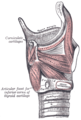
 Ligaments of the larynx. Posterior view. (Corniculate cartilage labeled at center right.) | |
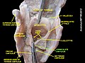
 The entrance to the larynx, viewed from behind. (Corniculate cartilage labeled at bottom right.) | |
| Details | |
| Identifiers | |
| Latin | cartilagines corniculatae |
| TA98 | A06.2.05.001 |
| TA2 | 997 |
| FMA | 55110 |
| Anatomical terminology [edit on Wikidata] | |
They are situated in the posterior parts of the aryepiglottic folds of mucous membrane, and are sometimes fused with the arytenoid cartilages.
Eponym edit
It is named by Giovanni Domenico Santorini.[1][2] The word "Corniculate" has a Latin root "cornu". Cornu means horn like projections. The projections of Corniculate cartilage look like "horns" hence the name.[3]
Additional images edit
-
 The cartilages of the larynx. Posterior view.
 -
 Laryngoscopic view of interior of larynx.
 -
 Muscles of larynx. Posterior view.
 -
 Muscles of larynx. Side view. Right lamina of thyroid cartilage removed.
 -
 Corniculate cartilages


References edit
![]() This article incorporates text in the public domain from page 1075 of the 20th edition of Gray's Anatomy (1918)
This article incorporates text in the public domain from page 1075 of the 20th edition of Gray's Anatomy (1918)
- ^ synd/3088 at Who Named It?
- ^ G. D. Santorini. Observationes anatomicae. Venetiis, apus J. B. Recurti, 1724; Leiden, 1939.
- ^ "Farlex free dictionary:Corniculate".
External links edit
- Atlas image: rsa3p8 at the University of Michigan Health System