


Summary
In human anatomy, the deltoid tuberosity is a rough, triangular[1] area on the anterolateral (front-side) surface of the middle of the humerus.[2] It is a site of attachment of deltoid muscle.[2]
| Deltoid tuberosity | |
|---|---|
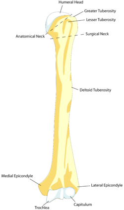 Left humerus. Anterior view. (Deltoideus labeled at center right.) | |
| Details | |
| Identifiers | |
| Latin | tuberositas deltoidea humeri |
| TA98 | A02.4.04.020 |
| TA2 | 1193 |
| FMA | 23418 |
| Anatomical terms of bone [edit on Wikidata] | |
Structure edit

Variation edit
The deltoid tuberosity has been reported as very prominent in less than 10% of people.[3]
Development edit
The deltoid tuberosity develops through endochondral ossification in a two-phase process.[4] The initiating signal is tendon-dependent, whilst the growth phase is muscle-dependent.[4]
Clinical significance edit
The deltoid tuberosity is at risk of avulsion fracture.[5] These fractures may be managed conservatively with rest.[5]
Other animals edit
In mammals, the humerus displays a wide morphological variation. The size and orientation of its functionally important features, including the deltoid tubercle, greater tubercle, and medial epicondyle, are pivotal to an animal's style of locomotion and habitat. In cursorial (running) animals such as the pronghorn, the deltoid tubercle is located about a quarter of the way down the shaft, which allows for rapid but relatively weak limb flexion and extension. In natatorial (swimming) animals such as the North American river otter, the tubercle is located nearly halfway down the shaft, which allows for powerful limb flexion and extension. The tuberosity can be very pronounced in fossorial (digging) animals, such as the mountain beaver.[6] It is very superficial in horses.[7]
See also edit
References edit
- ^ Gray, Henry (1918). Gray's Anatomy. ISBN 1-85958-018-1.
- ^ a b Feneis, Heinz (2000). Pocket Atlas of Human Anatomy (4th ed.). Thieme. p. 36. ISBN 3-13-511204-7.
- ^ Fink-Bennett D, Vicuna-Rios J. (1980). "The deltoid tuberosity--a potential pitfall (the "delta sign") in bone-scan interpretation: concise communication". The Journal of Nuclear Medicine. 21 (3): 211–212. PMID 7365512.
...in seven out of 100 scans reviewed.
- ^ a b Blitz, Einat; Viukov, Sergey; Sharir, Amnon; Shwartz, Yulia; Galloway, Jenna L.; Price, Brian A.; Johnson, Randy L.; Tabin, Clifford; Schweitzer, Ronen; Zelzer, Elazar (December 2009). "Bone ridge patterning during musculoskeletal assembly is mediated through SCX regulation of Bmp4 at the tendon-skeleton junction". Developmental Cell. 17 (6). Elsevier: 861–73. doi:10.1016/j.devcel.2009.10.010. PMC 3164485. PMID 20059955.
- ^ a b Nelson, Brad B.; Goodrich, Laurie R. (2014). "18 - Elbow and Shoulder". Equine Sports Medicine and Surgery (2nd ed.). Saunders Limited. pp. 343–365. doi:10.1016/B978-0-7020-4771-8.00018-1. ISBN 978-0-7020-4771-8.
- ^ Hall, Brian Keith (2007). Fins into limbs: evolution, development, and transformation. University of Chicago Press. p. 251. ISBN 978-0-226-31337-5. (Including an illustration of variation in mammalian humeri.)
- ^ Glass, Kati G.; Watkins, Jeffrey P. (2019). "Chapter 97 - Humerus". Equine Surgery (5th ed.). Saunders. pp. 1690–1699. doi:10.1016/B978-0-323-48420-6.00097-1. ISBN 978-0-323-48420-6.