


Summary
Dens invaginatus (DI), also known as tooth within a tooth, is a rare dental malformation and a developmental anomaly where there is an infolding of enamel into dentin. The prevalence of this condition is 0.3 - 10%,[1] affecting males more frequently than females. The condition presents in two forms, coronal involving tooth crown and radicular involving tooth root, with the former being more common.[2]
| Dens invaginatus | |
|---|---|
| Other names | Dens in dente, tooth within a tooth |
| Specialty | Dentistry |
DI is a malformation of teeth most likely resulting from an infolding of the dental papilla during tooth development or invagination of all layers of the enamel organ in dental papillae. Affected teeth show a deep infolding of enamel and dentin starting from the foramen coecum or even the tip of the cusps and which may extend deep into the root. Teeth most affected are maxillary lateral incisors (80%),[3] followed by maxillary canines (20%).[3] Bilateral occurrence is also seen (25%).[3]
Signs and symptoms edit
DI is often asymptomatic with the affected crown showing minimal external deformity. Individuals with an affected tooth may complain of their tooth having an abnormal shape such as being wider mesio-distally or bucco-lingually.[4]
Teeth affected by this condition are at a higher risk for developing caries and periradicular pathology.[1] The thin layer of the infolding enamel could be chipped off easily, providing entrance for microorganisms into the root canal. This can cause abscess formation and displacement of dental structures (i.e. teeth).[5] Early diagnosis and prevention is very important for maintaining tooth vitality. Clinical features such as incisal notching or pronounced talon cusp on lateral incisors could hit at DI and should be investigated with radiographs.[4]
Cause edit
Cause of DI is unclear. However, there are several theories:
Diagnosis edit
During clinical examination,[6] abnormally shaped tooth can be observed. Teeth with this condition can have a conical shape or deep pit on the lingual side or have an exaggerated talon cusp.
Although examination may reveal a fissure on the surface of anterior tooth, radiographic examination is the way.[7] On a periapical radiograph, the invagination lesion will appear as a radiolucent pocket. It is usually seen beneath the cingulum or incisal edge. Larger lesions can appear as fissures. A radio-opaque could be shown. Pulp may be involved and the root canal could have complex anatomy. Two periapical radiographs are often required to make sure that it is not a masked lesion.
Cone beam computed tomography[8][9] (CBCT) is useful in diagnosing DI. It provides clinicians a detailed 3D image and could aid treatment planning. Feasibility of root canal treatment or apical surgery or other procedures could be assessed.
Oehlers' classification edit
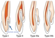
- Class I - Partial invagination. It is limited to the crown of tooth. The lesion does not extend pass the cementoenamel junction (CEJ) or the pulp.[1]
- Class II - Partial invagination. It extends beyond the crown and CEJ. Pulp may be involved but remain within the root anatomy. There is no communication of the lesion with periodontal ligament (PDL).[1]
- Class IIIa - Complete invagination. It extends through root and communicates with PDL. It usually does not involve the pulp but can cause anatomical malformation.[1]
- Class IIIb - Complete invagination. It extends through the root and communicates with PDL through apical foramen. Pulpal anatomy may not be directly involved but can cause disruption to the dental anatomy.[1]
Histology edit
Management edit
References edit
- ^ a b c d e f g h i j A. Gallacher, R. Ali & S. Bhakta (15 August 2016). "Dens invaginatus: diagnosis and management strategies". British Dental Journal. 221 (7): 383–387. doi:10.1038/sj.bdj.2016.724. PMID 27713460. S2CID 12853879.
- ^ Alves dos Santos, Guilherme Nilson; Sousa-Neto, Manoel Damião; Assis, Helena Cristina; Lopes-Olhê, Fabiane Carneiro; Faria-e-Silva, André L.; Oliveira, Matheus L.; Mazzi-Chaves, Jardel Francisco; Candemil, Amanda Pelegrin (July 2023). "Prevalence and morphological analysis of dens invaginatus in anterior teeth using cone beam computed tomography: A systematic review and meta-analysis". Archives of Oral Biology. 151: 105715. doi:10.1016/j.archoralbio.2023.105715. ISSN 0003-9969.
- ^ a b c Hakan Çolak, Enes Tan,Bahadır Uğur Aylıkçı, Recep Uzgur, Mustafa Turkal, and Mehmet Mustafa Hamidi (29 June 2012). "Radiographic Study of the Prevalence of Dens Invaginatus in a Sample Set of Turkish Dental Patients". Journal of Clinical Imaging Science. 2: 34. doi:10.4103/2156-7514.97755. PMC 3424816. PMID 22919548.
{{cite journal}}: CS1 maint: multiple names: authors list (link) - ^ a b Gallacher, A.; Ali, R.; Bhakta, S. (2016-10-07). "Dens invaginatus: diagnosis and management strategies". British Dental Journal. 221 (7): 383–387. doi:10.1038/sj.bdj.2016.724. ISSN 0007-0610.
- ^ a b c d e f g h "What is dens invaginatus or dens in dente?". 3 Feb 2018.
- ^ a b Schmitz MS, Montagner F, Flores CB, Morari VH, Quesada GA, Gomes BP (June 2010). "Management of dens invaginatus type I and open apex: report of three cases". Journal of Endodontics. 36 (6): 1079–1085. doi:10.1016/j.joen.2010.02.002. PMID 20478470.
- ^ Radicular Dens Invaginatus: Report of a Rare Case https://www.hindawi.com/journals/crid/2012/871937/
- ^ a b Pushpak Narayana; Gary R Hartwell; Robert Wallace; Umadevi P Nair (August 2012). "Endodontic Clinical Management of a Dens Invaginatus Case by Using a Unique Treatment Approach: A Case Report". Journal of Endodontics. 38 (8): 1145–8. doi:10.1016/j.joen.2012.04.020. PMID 22794224.
- ^ Álvaro Zubizarreta Macho; Alberto Ferreiroa; Cristina Rico-Romano; Luis Óscar Alonso-Ezpeleta; Jesús Mena-Álvarez (April 2015). "Diagnosis and endodontic treatment of type II dens invaginatus by using cone-beam computed tomography and splint guides for cavity access". The Journal of the American Dental Association. 146 (4): 266–70. doi:10.1016/j.adaj.2014.11.021. PMID 25819658.
- ^ a b c d Piattelli A, Trisi P (1993). "Dens invaginatus: a histological study of undermineralized material". Dental Traumatology. 9 (5): 191–195. doi:10.1111/j.1600-9657.1993.tb00273.x.
- ^ a b Satyaranjan Mishra, Lora Mishra, and Sujit Ranjan Sahoo (Nov 2012). "A Type III Dens Invaginatus with Unusual Helical CT and Histologic Findings: A Case Report". Journal of Clinical and Diagnostic Research.
{{cite journal}}: CS1 maint: multiple names: authors list (link) - ^ Harleen Kumar; Muna Al-Ali; Peter Parashos; David J Manton (May 2014). "Management of 2 Teeth Diagnosed with Dens Invaginatus with Regenerative Endodontics and Apexification in the Same Patient: A Case Report and Review". Journal of Endodontics. 40 (5): 725–31. doi:10.1016/j.joen.2013.10.030. PMID 24767572.