


Summary
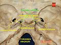
The foramen lacerum (Latin: lacerated piercing) is a triangular hole in the base of skull. It is located between the sphenoid bone, the apex of the petrous part of the temporal bone, and the basilar part of the occipital bone.
| Foramen lacerum | |
|---|---|
 | |
| Details | |
| System | skeletal |
| Parts | temporal bone, sphenoid bone, occipital bone |
| Identifiers | |
| Latin | foramen lacerum |
| TA98 | A02.1.00.055 |
| TA2 | 459 |
| FMA | 54809 |
| Anatomical terminology [edit on Wikidata] | |
Structure edit
The foramen lacerum (Latin: lacerated piercing) is a triangular hole in the base of skull. It is located between 3 bones:
- sphenoid bone (forming the anterior border)[1]: 776
- apex of petrous part of temporal bone (forming the posterolateral border)[1]: 776 [2]
- basilar part of occipital bone (forming the posteromedial border)[1]: 776
It is the junction point of 3 sutures of the skull:
- petroclival (petrooccipital) suture[1]: 776 [3]
- sphenopetrosal suture[1]: 776
- sphenooccipital suture[3]
Contents edit
Structures passing through the foramen lacerum include:
- greater petrosal nerve and deep petrosal nerve which merge within the foramen to form the nerve of the pterygoid canal[4]
- nerve of the pterygoid canal[citation needed]
- artery of the pterygoid canal[citation needed]
- recurrent artery of the foramen lacerum (supplies the internal carotid plexus)[5]
- emissary veins (connecting extracranial pterygoid plexus with the intracranial cavernous sinus)[6]
- one of the terminal branches of the ascending pharyngeal artery[citation needed]
Relations edit
It is situated anteromedially to the carotid canal.[1]: 776
The internal carotid artery passes from the carotid canal in the base of the skull, emerging and coursing superior to foramen lacerum as it exits the carotid canal; the internal carotid artery does not travel through foramen lacerum (the segment of the internal carotid artery that travels superior to the foramen lacerum is called the lacerum segment).[7]
Development edit
The foramen lacerum fills with cartilage after birth.[1]: 776
Clinical significance edit
The foramen lacerum has been described as a portal of entry into the cranium for tumours, including nasopharyngeal carcinoma, juvenile angiofibroma, adenoid cystic carcinoma, malignant melanoma, and lymphoma.[8][9]
History edit
The first recorded mention of the foramen lacerum was by anatomist Wenzel Gruber in 1869.[10][8] Study of the foramen has been neglected for many years because of the small role it plays in intracranial surgery.[8]
Additional images edit
-
 Foramen lacerum

References edit
- ^ a b c d e f g Drake, Richard L.; Vogl, Wayne; Tibbitts, Adam W.M. Mitchell; illustrations by Richard; Richardson, Paul (2005). Gray's anatomy for students. Philadelphia: Elsevier/Churchill Livingstone. ISBN 978-0-8089-2306-0.
- ^ Chae, Ricky; Rubio, Roberto Rodriguez (2020). "14 - Anatomy of petrous face". Handbook of Clinical Neurology. Vol. 170. Elsevier. pp. 143–156. doi:10.1016/B978-0-12-822198-3.00036-7. ISBN 978-0-12-822198-3. ISSN 0072-9752. PMID 32586486. S2CID 220077819.
- ^ a b Netter, Frank H.; Norton, Neil S. (2011). Netter's Head and Neck Anatomy for Dentistry (2nd ed.). Elsevier, Saunders. pp. 47–50.
- ^ Goosmann, Madeline M.; Dalvin, Mark (2023), "Anatomy, Head and Neck, Deep Petrosal Nerve", StatPearls, Treasure Island (FL): StatPearls Publishing, PMID 30521238, retrieved 2023-07-31
- ^ Seker, Askin; Martins, Carolina; Rhoton, Albert L. (2010). "2 - Meningeal Anatomy". Meningiomas. Saunders. pp. 11–51. doi:10.1016/B978-1-4160-5654-6.00002-7. ISBN 978-1-4160-5654-6.
{{cite book}}: CS1 maint: location missing publisher (link) - ^ Sinnatamby, Chummy S. (2011). Last's Anatomy (12th ed.). Elsevier Australia. p. 364. ISBN 978-0-7295-3752-0.
- ^ Tubbs, R. Shane; Shoja, Mohammadali M.; Loukas, Marios (25 April 2016). Bergman's Comprehensive Encyclopedia of Human Anatomic Variation. John Wiley & Sons. p. 450. ISBN 9781118430279.
- ^ a b c Tauber, M; van Loveren, HR; Jallo, G; Romano, A; Keller, JT (February 1999). "The enigmatic foramen lacerum". Neurosurgery. 44 (2): 386–91, discussion 391-3. doi:10.1097/00006123-199902000-00083. PMID 9932893.
- ^ Christodouleas, Boris Hristov, Steven H. Lin, John P. (2010). Radiation oncology : a question-based review. Philadelphia, Pa.: Lippincott Williams & Wilkins. p. 138. ISBN 978-1608314447.
{{cite book}}: CS1 maint: multiple names: authors list (link) - ^ Gruber, Wenzel (1869). Beitrage Zur Anatomie Des Schadelgrundes. ISBN 9781162306223.
External links edit
- Anatomy figure: 22:5b-10 at Human Anatomy Online, SUNY Downstate Medical Center - "Internal view of skull."
- Photo of model at Waynesburg College skeleton/foramenlacerum
- cranialnerves at The Anatomy Lesson by Wesley Norman (Georgetown University) (VII)
- "Anatomy diagram: 34257.000-1". Roche Lexicon - illustrated navigator. Elsevier. Archived from the original on 2012-07-22.
- Image at ucsd.edu Archived 2021-10-21 at the Wayback Machine