


Summary
The lateral pectoral nerve (also known as the lateral anterior thoracic nerve) arises from the lateral cord of the brachial plexus,[1][2] and through it from the C5-7.[1][2]
| Lateral pectoral nerve | |
|---|---|
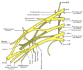
 Nerves of the left upper extremity. (Lateral anterior thoracic visible in upper right.) | |
| Details | |
| From | Lateral cord |
| Innervates | pectoralis major |
| Identifiers | |
| Latin | nervus pectoralis lateralis |
| TA98 | A14.2.03.018 |
| TA2 | 6419 |
| FMA | 65296 |
| Anatomical terms of neuroanatomy [edit on Wikidata] | |
It passes across the axillary artery and vein,[3] pierces the clavipectoral (coracoclavicular) fascia, and enters the deep surface of the pectoralis major to innervate it.[1][4]
Function edit
The lateral pectoral nerve provides motor innervation to the pectoralis major muscle.[2][5]
Although this nerve is described as mostly motor, it also has been considered to carry proprioceptive and nociceptive fibers. It arises either from the lateral cord or directly from the anterior divisions of the upper and middle trunks of the brachial plexus. This is unlike the medial pectoral nerve, which derives from the medial cord (or directly from the anterior division of the lower trunk). It splits into four to seven branches that pierce the clavipectoral fascia to innervate the entire pectoralis major or its superior portion.
The medial and lateral pectoral nerves form a connection, around the axillary artery, called the ansa pectoralis. The lateral pectoral nerve has been described as double, while the medial pectoral nerve has been described as single.[6]
Clinical significance edit
Postoperative Care edit
The lateral pectoral nerve is important in the pain response after breast augmentation and mastectomy, and especially in breast implant surgery, when the implant is inserted by the subpectoral route. The pectoral nerves can be anesthetized (blocked) intraoperatively by the surgeon under direct vision by three injections - one to block the medial pectoral nerve, the second to block the perforating branches of the medial pectoral nerve, and the third to block the lateral pectoral nerve. An ultrasound-guided pectoral nerve block can also be performed preventively before the operation by an anesthesiologist, experienced in regional anesthesia. It is safe and relies on ultrasound imaging to localize the pectoralis major and minor muscles, the presumed course of the pectoral nerves and the optimal spread of the local anesthetic.[7]
Nerve Block edit
Blockade of the lateral pectoral nerve is helpful in cases such as shoulder dislocation and other orthopedic procedures, involving the shoulder. Spasms of the pectoralis major muscle and resulting severe pain (acute or chronic) may be reduced by pectoral nerve block or neuromuscular relaxation. Decreasing the pectoral muscle tone intraoperatively by neuromuscular relaxation (paralytic agents) or by a nerve block (local anesthetic injection), can facilitate better cosmetic results during breast augmentation or post-mastectomy breast implantation. “The skin projection point of the neurovascular bundle (NVB) represents the denervation point (DP).” The NVB (thoracoacromial artery and vein, plus the lateral pectoral nerve) may be the guide for local anesthetic applications in order to achieve pectoral muscle denervation. “Routine botulinum toxin infiltration of the chest wall musculature at the time of mastectomy and immediate reconstruction… would paralyze the muscles and reduce the postoperative pain caused by muscle spasm.”[8]
Damage edit
The lateral pectoral nerve can be damaged, particularly during surgery, by cauterisation and avulsion.[4] Trauma to the brachial plexus can cut off innervation to the lateral pectoral nerve.[9]
See also edit
References edit
![]() This article incorporates text in the public domain from page 933 of the 20th edition of Gray's Anatomy (1918)
This article incorporates text in the public domain from page 933 of the 20th edition of Gray's Anatomy (1918)
- ^ a b c Kahn, Elyne; Yang, Lynda J. -S. (1 January 2015), Tubbs, R. Shane; Rizk, Elias; Shoja, Mohammadali M.; Loukas, Marios (eds.), "Chapter 40 - Anatomy of the Lateral Cord and Its Branches", Nerves and Nerve Injuries, San Diego: Academic Press, pp. 547–551, doi:10.1016/b978-0-12-410390-0.00042-1, ISBN 978-0-12-410390-0, retrieved 19 October 2020
- ^ a b c Baur, Dale A.; Horan, Michael P.; Rodriguez, Juan C. (1 January 2012), Bagheri, Shahrokh C.; Bell, R. Bryan; Khan, Husain Ali (eds.), "Chapter 68 - The Pectoralis Major Myocutaneous Flap", Current Therapy In Oral and Maxillofacial Surgery, Saint Louis: W.B. Saunders, pp. 566–572, doi:10.1016/b978-1-4160-2527-6.00068-2, ISBN 978-1-4160-2527-6, retrieved 26 October 2020
- ^ Rea, Paul (1 January 2015), Rea, Paul (ed.), "Chapter 2 - Upper Limb Nerve Supply", Essential Clinically Applied Anatomy of the Peripheral Nervous System in the Limbs, Academic Press, pp. 41–100, doi:10.1016/b978-0-12-803062-2.00002-4, ISBN 978-0-12-803062-2, retrieved 26 October 2020
- ^ a b Willey, Shawna C.; Feldman, Elizabeth D. (1 January 2009), Evans, Stephen R. T. (ed.), "Chapter 46 - Mastectomy", Surgical Pitfalls, Philadelphia: W.B. Saunders, pp. 475–487, doi:10.1016/b978-141602951-9.50058-x, ISBN 978-1-4160-2951-9, retrieved 26 October 2020
- ^ Wei, William Ignace; Chan, Yu-wai (1 January 2009), Wei, Fu-Chan; Mardini, Samir (eds.), "CHAPTER 17 - Pectoralis major flap", Flaps and Reconstructive Surgery, Edinburgh: W.B. Saunders, pp. 175–192, doi:10.1016/b978-0-7216-0519-7.00017-4, ISBN 978-0-7216-0519-7, retrieved 26 October 2020
- ^ Porzionato, Macchi; Stecco (2012). "Surgical Anatomy of the Pectoral Nerves and the Pectoral Musculature". Clinical Anatomy. 25 (5): 559–575. doi:10.1002/ca.21301. PMID 22125052. S2CID 22125503.
- ^ Desroches, Jean; Grabs (2013). "). "Selective Ultrasound Guided Pectoral Nerve Targeting in Breast Augmentation: How to Spare the Brachial Plexus Cords?"". Clinical Anatomy. 26 (1): 49–55. doi:10.1002/ca.22117. PMID 22730005. S2CID 26711722.
- ^ Titiz, Izzet; Ozel; Ozel; Toros; Marur; Yildirim; Erdogdu; Kara (19 August 2010). "Denervation Point for Neuromuscular Blockade on Lateral Pectoral Nerves: A Cadaver Study". Surgical and Radiologic Anatomy. 33 (2): 105–108. doi:10.1007/s00276-010-0712-7. PMID 20721553. S2CID 6959180.
- ^ Larsen, Mikko; Bishop, Allen T.; Shin, Alexander Y. (1 January 2018), Morrey, Bernard F.; Sanchez-Sotelo, Joaquin; Morrey, Mark E. (eds.), "117 - Flaccid Dysfunction", Morrey's the Elbow and its Disorders (Fifth Edition), Philadelphia: Content Repository Only!, pp. 1078–1098, ISBN 978-0-323-34169-1, retrieved 26 October 2020
Additional images edit
-
 Plan of brachial plexus.
 -
 Brachial plexus
 -
 Schema of brachial plexus with course of spinal nerves shown.

External links edit
- Anatomy photo:05:st-0506 at the SUNY Downstate Medical Center
- EatonHand ner-014
- Photo at mun.ca
- MedEd at Loyola grossanatomy/dissector/labs/ue/pect_scap/p2_1.html