


Summary
The levator veli palatini (/lɪˈveɪtər ˈviːlaɪ ˌpæləˈtaɪnaɪ/) is a muscle of the soft palate and pharynx. It is innervated by the vagus nerve (cranial nerve X) via its pharyngeal plexus. During swallowing, it contracts, elevating the soft palate to help prevent food from entering the nasopharynx.
| Levator veli palatini | |
|---|---|
 Dissection of the muscles of the palate from behind. (Caption for Levator veli palatini visible at right, second from the top.) | |
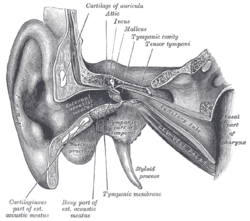 External and middle ear, opened from the front. Right side. (Levator veli palatini visible at bottom right.) | |
| Details | |
| Origin | temporal bone, Eustachian tube |
| Insertion | palatine aponeurosis |
| Artery | facial artery |
| Nerve | Pharyngeal Branch of Vagus (CN X) |
| Actions | elevates soft palate |
| Identifiers | |
| Latin | musculus levator veli palatini |
| TA98 | A05.2.01.102 |
| TA2 | 2128 |
| FMA | 46727 |
| Anatomical terms of muscle [edit on Wikidata] | |
Structure edit
The levator veli palatini muscle occurs in the soft palate of the mouth.[1] It forms a sling superior and immediately posterior to the palatine aponeurosis.[2]
Origin edit
The primary site of origin of the muscle is a quadrangular roughened area upon the medial extremity of the inferior aspect of the petrous part of the temporal bone; here, the muscle arises by a small tendon.[2]
Additional fibres of the muscle arise from the inferior aspect of the cartilaginous part of pharyngotympanic tube, and the vaginal process of sphenoid bone.[2]
Insertion edit
In the medial third of the soft palate, its fibers spread out between the two strands of the palatoglossus muscle to attach to the superior surface of the palatine aponeurosis and intermingle with fibres of its contralateral partner.[2]
Nerve supply edit
The levator veli palatini muscle receives motor innervation from the vagus nerve (CN X)[citation needed] via the pharyngeal plexus.[2]
Relations edit
During its course from its origin to its insertion, the muscle passes medial to the superior margin of the superior pharyngeal constrictor muscle.[2]
It lies lateral to the choana.[citation needed]
Actions/movements edit
The primary action of the muscle is to elevate and draw posterior-ward the nearly vertical posterior portion of the soft palate; thereby, the soft palate is brought into contact with the posterior wall of the pharynx, thus creating a barrier between the nasopharynx and oropharynx.[2]
Additionally, the muscle draws the lateral walls of the nasopharynx posteromedially, thus narrowing the nasopharynx.[2]
Function edit
The levator veli palatini muscle elevates the soft palate during swallowing. This helps to prevent food from entering the nasopharynx. Its action may be slightly slower than its partner, the tensor veli palatini muscle.[1]
It has little to no effect on the pharyngotympanic tube.[2]
Additional images edit
-
 Left temporal bone. Inferior surface.

References edit
![]() This article incorporates text in the public domain from page 1139 of the 20th edition of Gray's Anatomy (1918)
This article incorporates text in the public domain from page 1139 of the 20th edition of Gray's Anatomy (1918)
- ^ a b Ishijima, Ken; Sando, Isamu; Miura, Makoto; Balaban, Carey D.; Takasaki, Kenji (2002-06-01). "Functional Anatomy of Levator Veli Palatini Muscle and Tensor Veli Palatini Muscle in Association with Eustachian Tube Cartilage". Annals of Otology, Rhinology & Laryngology. 111 (6): 530–536. doi:10.1177/000348940211100609. ISSN 0003-4894. PMID 12090709. S2CID 36727539.
- ^ a b c d e f g h i Standring, Susan (2020). Gray's Anatomy: The Anatomical Basis of Clinical Practice (42th ed.). New York. p. 709. ISBN 978-0-7020-7707-4. OCLC 1201341621.
{{cite book}}: CS1 maint: location missing publisher (link)
External links edit
- "Anatomy diagram: 25420.000-1". Roche Lexicon - illustrated navigator. Elsevier. Archived from the original on 2014-01-01.