


Summary
Magnetic resonance imaging of the brain uses magnetic resonance imaging (MRI) to produce high quality two-dimensional or three-dimensional images of the brain and brainstem as well as the cerebellum without the use of ionizing radiation (X-rays) or radioactive tracers.
| MRI of Brain | |
|---|---|
 Cross-sectional T1-weighted MRI of a healthy human brain acquired with an ultra high-field MR of 7 Tesla field strength | |
| ICD-10-PCS | B030ZZZ |
| ICD-9-CM | 88.91 |
| OPS-301 code | 3-800, 3-820 |
History edit
The first MR images of a human brain were obtained in 1978 by two groups of researchers at EMI Laboratories led by Ian Robert Young and Hugh Clow.[1] In 1986, Charles L. Dumoulin and Howard R. Hart at General Electric developed MR angiography,[2] and Denis Le Bihan obtained the first images and later patented diffusion MRI.[3] In 1988, Arno Villringer and colleagues demonstrated that susceptibility contrast agents may be employed in perfusion MRI.[4] In 1990, Seiji Ogawa at AT&T Bell labs recognized that oxygen-depleted blood with dHb was attracted to a magnetic field, and discovered the technique that underlies Functional Magnetic Resonance Imaging (fMRI).[5]

In the early 1980s to the early 1990s, 'Jedi' helmets, inspired by the 'Return of the Jedi' Star Wars film, were sometimes worn by children in order to obtain good image quality. The copper coils of the helmet were used as a radio aerial to detect the signals while the 'Jedi' association encouraged children to wear the helmets and not be frightened by the procedure. These helmets were no longer needed as MR scanners improved.
In the early 1990s, Peter Basser and Le Bihan, working at NIH, and Aaron Filler, Franklyn Howe, and colleagues developed diffusion tensor imaging (DTI).[6][7][8][9] Joseph Hajnal, Young and Graeme Bydder described the use of FLAIR pulse sequence to demonstrate high signal regions in normal white matter in 1992.[10] In the same year, John Detre, Alan P. Koretsky and coworkers developed arterial spin labeling.[11] In 1997, Jürgen R. Reichenbach, E. Mark Haacke and coworkers at Washington University in St. Louis developed Susceptibility weighted imaging.[12]
The first study of the human brain at 3.0 T was published in 1994,[13] and in 1998 at 8 T.[14] Studies of the human brain have been performed at 9.4 T (2006)[15] and up to 10.5 T (2019).[16]
Paul Lauterbur and Sir Peter Mansfield were awarded the 2003 Nobel Prize in Physiology or Medicine for their discoveries concerning MRI.

The record for the highest spatial resolution of a whole intact brain (postmortem) is 100 microns, from Massachusetts General Hospital. The data was published in Scientific Data on 30 October 2019.[17][18]
Applications edit
One advantage of MRI of the brain over computed tomography of the head is better tissue contrast,[19] and it has fewer artifacts than CT when viewing the brainstem. MRI is also superior for pituitary imaging.[20] It may however be less effective at identifying early cerebritis.[21]
In the case of a concussion, an MRI should be avoided unless there are progressive neurological symptoms, focal neurological findings or concern of skull fracture on exam.[22] In the analysis of a concussion, measurements of Fractional Anisotropy, Mean Diffusivity, Cerebral Blood Flow, and Global Connectivity can be taken to observe the pathophysiological mechanisms being made while in recovery.[23]
In analysis of the fetal brain, MRI provides more information about gyration than ultrasound.[24]
MRI is sensitive for the detection of brain abscess.[25]
A number of different imaging modalities or sequences can be used with imaging the nervous system:
- T1-weighted (T1W) images: Cerebrospinal fluid is dark. T1-weighted images are useful for visualizing normal anatomy.
- T2-weighted (T2W) images: CSF is light, but fat (and thus white matter) is darker than with T1. T2-weighted images are useful for visualizing pathology.[26]
- Diffusion-weighted images (DWI): DWI uses the diffusion of water molecules to generate contrast in MR images.
- Proton density (PD) images: CSF has a relatively high level of protons, making CSF appear bright. Gray matter is brighter than white matter.[27]

- Fluid attenuation inversion recovery (FLAIR): useful for evaluation of white matter plaques near the ventricles.[28] It is useful in identifying demyelination.[29]
Diagnostic Usage edit
MRI of the brain and head has multiple diagnostic usages, including identifying aneurysms, strokes, tumors and other brain injury.[30] In many diseases, such as Parkinson's or Alzheimer's, MRI is useful to help differentially diagnose against other diseases.[31][32] On the topic of diagnosis, MRI data has been used with deep learning networks to identify brain tumors.[33]
See also edit
Gallery edit
-
 Brain regions on T1 MRI
 -
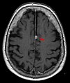 T1 (note CSF is dark) with contrast (arrow pointing to meningioma of the falx)
 -
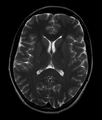 Normal axial T2-weighted MR image of the brain
 -
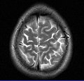 MRI image of the surface of the brain.


References edit
- ^ "Britain's brains produce first NMR scans". New Scientist: 588. 1978.
- ^ "Blood-flow checker". Popular Science: 12. 1987.
- ^ Le Bihan D, Breton E (1987). "Method to Measure the Molecular Diffusion and/or Perfusion Parameters of Live Tissue". US Patent # 4,809,701.
- ^ Villringer A, Rosen BR, Belliveau JW, Ackerman JL, Lauffer RB, Buxton RB, Chao YS, Wedeen VJ, Brady TJ (February 1988). "Dynamic imaging with lanthanide chelates in normal brain: contrast due to magnetic susceptibility effects". Magnetic Resonance in Medicine. 6 (2): 164–74. doi:10.1002/mrm.1910060205. PMID 3367774. S2CID 41228095.
- ^ Faro SH, Mohamed FB (2010-01-15). Bold fMRI. a guide to functional imaging for neuroscientists. Springer. ISBN 978-1-4419-1328-9. Retrieved 10 June 2015.
- ^ Howe FA, Filler AG, Bell BA, Griffiths JR (December 1992). "Magnetic resonance neurography". Magnetic Resonance in Medicine. 28 (2): 328–38. doi:10.1002/mrm.1910280215. PMID 1461131. S2CID 36417513.
- ^ Filler AG, Howe FA, Hayes CE, Kliot M, Winn HR, Bell BA, Griffiths JR, Tsuruda JS (March 1993). "Magnetic resonance neurography". Lancet. 341 (8846): 659–61. doi:10.1016/0140-6736(93)90422-d. PMID 8095572. S2CID 24795253.
- ^ Filler A (October 2009). "Magnetic resonance neurography and diffusion tensor imaging: origins, history, and clinical impact of the first 50,000 cases with an assessment of efficacy and utility in a prospective 5000-patient study group". Neurosurgery. 65 (4 Suppl): A29-43. doi:10.1227/01.neu.0000351279.78110.00. PMC 2924821. PMID 19927075.
- ^ Basser PJ (2010). "Invention and Development of Diffusion Tensor MRI (DT-MRI or DTI) at the NIH". Diffusion MRI. Oxford University Press. pp. 730–740. doi:10.1093/med/9780195369779.003.0047. ISBN 9780195369779.
- ^ Hajnal JV, De Coene B, Lewis PD, Baudouin CJ, Cowan FM, Pennock JM, Young IR, Bydder GM (July 1992). "High signal regions in normal white matter shown by heavily T2-weighted CSF nulled IR sequences". Journal of Computer Assisted Tomography. 16 (4): 506–13. doi:10.1097/00004728-199207000-00002. PMID 1629405. S2CID 42727826.
- ^ Koretsky AP (August 2012). "Early development of arterial spin labeling to measure regional brain blood flow by MRI". NeuroImage. 62 (2): 602–7. doi:10.1016/j.neuroimage.2012.01.005. PMC 4199083. PMID 22245338.
- ^ Reichenbach JR, Venkatesan R, Schillinger DJ, Kido DK, Haacke EM (July 1997). "Small vessels in the human brain: MR venography with deoxyhemoglobin as an intrinsic contrast agent". Radiology. 204 (1): 272–7. doi:10.1148/radiology.204.1.9205259. PMID 9205259.
- ^ Mansfield P, Coxon R, Glover P (May 1994). "Echo-planar imaging of the brain at 3.0 T: first normal volunteer results". Journal of Computer Assisted Tomography. 18 (3): 339–43. doi:10.1097/00004728-199405000-00001. PMID 8188896. S2CID 20221062.
- ^ Robitaille PM, Abduljalil AM, Kangarlu A, Zhang X, Yu Y, Burgess R, Bair S, Noa P, Yang L, Zhu H, Palmer B, Jiang Z, Chakeres DM, Spigos D (October 1998). "Human magnetic resonance imaging at 8 T". NMR in Biomedicine. 11 (6): 263–5. doi:10.1002/(SICI)1099-1492(199810)11:6<263::AID-NBM549>3.0.CO;2-0. PMID 9802467. S2CID 41305659.
- ^ Vaughan T; DelaBarre L; Snyder C; Tian J; Akgun C; Shrivastava D; Liu W; Olson C; Adriany G; et al. (December 2006). "9.4T human MRI: preliminary results". Magn Reson Med. 56 (6): 1274–82. doi:10.1002/mrm.21073. PMC 4406343. PMID 17075852.
- ^ Sadeghi‐Tarakameh, Alireza; DelaBarre, Lance; Lagore, Russell L.; Torrado‐Carvajal, Angel; Wu, Xiaoping; Grant, Andrea; Adriany, Gregor; Metzger, Gregory J.; Van de Moortele, Pierre‐Francois; Ugurbil, Kamil; Atalar, Ergin (2019-11-21). "In vivo human head MRI at 10.5T: A radiofrequency safety study and preliminary imaging results". Magnetic Resonance in Medicine. 84 (1): 484–496. doi:10.1002/mrm.28093. hdl:11693/53263. ISSN 0740-3194. PMC 7695227. PMID 31751499. S2CID 208226414.
- ^ "100-Hour-Long MRI of Human Brain Produces Most Detailed 3D Images Yet". 10 July 2019.
- ^ "Team publishes on highest resolution brain MRI scan".
- ^ Ebel KD, Benz-Bohm G (1999). Differential diagnosis in pediatric radiology. Thieme. pp. 538–. ISBN 978-3-13-108131-5. Retrieved 18 July 2011.
- ^ Bradley WG, Brant-Zawadzki M, Cambray-Forker J (2001-01-15). MRI of the brain. Surendra Kumar. ISBN 978-0-7817-2568-2. Retrieved 24 July 2011.
- ^ Roos KL, Tunkel AR (2010). Bacterial infections of the central nervous system. Elsevier Health Sciences. pp. 69–. ISBN 978-0-444-52015-9. Retrieved 18 July 2011.
- ^ American Medical Society for Sports Medicine (24 April 2014), "Five Things Physicians and Patients Should Question", Choosing Wisely: an initiative of the ABIM Foundation, American Medical Society for Sports Medicine, retrieved 29 July 2014
- ^ Churchill Nathan W., Hutchison Michael G., Richards Doug, Leung General, Graham Simon J., Schweizer Tom A. (2017). "The first week after concussion: Blood flow, brain function and white matter microstructure". NeuroImage: Clinical. 14: 480–489. doi:10.1016/j.nicl.2017.02.015. PMC 5334547. PMID 28280686.
{{cite journal}}: CS1 maint: multiple names: authors list (link) - ^ Garel C (2004). MRI of the fetal brain: normal development and cerebral pathologies. Springer. ISBN 978-3-540-40747-8. Retrieved 24 July 2011.
- ^ Rath, Tanya J.; Hughes, Marion; Arabi, Mohammad; Shah, Gaurang V. (2012). "Imaging of Cerebritis, Encephalitis, and Brain Abscess". Neuroimaging Clinics of North America. 22 (4). Elsevier BV: 585–607. doi:10.1016/j.nic.2012.04.002. ISSN 1052-5149. PMID 23122258.
- ^ Butler P, Mitchell AW, Ellis H (2007-11-19). Applied Radiological Anatomy for Medical Students. Cambridge University Press. pp. 12–. ISBN 978-0-521-81939-8. Retrieved 18 July 2011.
- ^ Tofts, Paul (2005-09-01). Quantitative MRI of the Brain: Measuring Changes Caused by Disease. John Wiley and Sons. pp. 86–. ISBN 978-0-470-86949-9. Retrieved 18 July 2011.
- ^ Chowdhury R, Wilson I, Rofe C, Lloyd-Jones G (2010-04-19). Radiology at a Glance. John Wiley and Sons. pp. 95–. ISBN 978-1-4051-9220-0. Retrieved 18 July 2011.
- ^ Granacher RP (2007-12-20). Traumatic brain injury: methods for clinical and forensic neuropsychiatric assessment. CRC Press. pp. 247–. ISBN 978-0-8493-8138-6. Retrieved 18 July 2011.
- ^ "MRI - Mayo Clinic". www.mayoclinic.org. Retrieved 2023-12-22.
- ^ Heim, Beatrice; Krismer, Florian; De Marzi, Roberto; Seppi, Klaus (2017-08-01). "Magnetic resonance imaging for the diagnosis of Parkinson's disease". Journal of Neural Transmission. 124 (8): 915–964. doi:10.1007/s00702-017-1717-8. ISSN 1435-1463. PMC 5514207. PMID 28378231.
- ^ Frisoni, Giovanni B.; Fox, Nick C.; Jack, Clifford R.; Scheltens, Philip; Thompson, Paul M. (February 2010). "The clinical use of structural MRI in Alzheimer disease". Nature Reviews Neurology. 6 (2): 67–77. doi:10.1038/nrneurol.2009.215. ISSN 1759-4766. PMC 2938772.
- ^ Segato, Alice; Marzullo, Aldo; Calimeri, Francesco; De Momi, Elena (2020-12-01). "Artificial intelligence for brain diseases: A systematic review". APL Bioengineering. 4 (4). AIP Publishing: 041503. doi:10.1063/5.0011697. ISSN 2473-2877. PMC 7556883. PMID 33094213.