


Summary
Nanodiamonds, or diamond nanoparticles, are diamonds with a size below 100 nanometers.[2] They can be produced by impact events such as an explosion or meteoritic impacts. Because of their inexpensive, large-scale synthesis, potential for surface functionalization, and high biocompatibility, nanodiamonds are widely investigated as a potential material in biological and electronic applications and quantum engineering.[3][4]


History edit
In 1963, Soviet scientists at the All-Union Research Institute of Technical Physics noticed that nanodiamonds were created by nuclear explosions that used carbon-based trigger explosives.[3][5]
Structure and composition edit
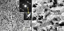
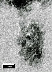
There are three main aspects in the structure of diamond nanoparticles to be considered: the overall shape, the core, and the surface. Through multiple diffraction experiments, it has been determined that the overall shape of diamond nanoparticles is either spherical or elliptical. At the core of diamond nanoparticles lies a diamond cage, which is composed mainly of carbons.[6] While the core closely resemble the structure of a diamond, the surface of diamond nanoparticles actually resemble the structure of graphite. A recent study shows that the surface consists mainly of carbons, with high amounts of phenols, pyrones, and sulfonic acid, as well as carboxylic acid groups, hydroxyl groups, and epoxide groups, though in lesser amounts.[7] Occasionally, defects such as nitrogen-vacancy centers can be found in the structure of diamond nanoparticles. 15N NMR research confirms presence of such defects.[8] A recent study shows that the frequency of nitrogen-vacancy centers decreases with the size of diamond nanoparticles.[9]
 |
 |
 |
Production methods edit

Other than explosions, methods of synthesis include hydrothermal synthesis, ion bombardment, laser bombardment, microwave plasma chemical vapor deposition techniques, ultrasound synthesis,[10] and electrochemical synthesis.[11] In addition, the decomposition of graphitic C3N4 under high pressure and high temperature yields large quantities of high purity diamond nanoparticles.[12] However, detonation synthesis of nanodiamonds has become the industry standard in the commercial production of nanodiamonds: the most commonly utilized explosives being mixtures of trinitrotoluene and hexogen or octogen. Detonation is often performed in a sealed, oxygen-free, stainless steel chamber and yields a mixture of nanodiamonds averaging 5 nm and other graphitic compounds.[13] In detonation synthesis, nanodiamonds form under pressures greater than 15 GPa and temperatures greater than 3000K in the absence of oxygen to prevent the oxidation of diamond nanoparticles.[13] The rapid cooling of the system increases nanodiamond yields as diamond remains the most stable phase under such conditions. Detonation synthesis utilizes gas-based and liquid-based coolants such as argon and water, water-based foams, and ice.[13] Because detonation synthesis results in a mix of nanodiamond particles and other graphitic carbon forms, extensive cleaning methods must be employed to rid the mixture of impurities. In general, gaseous ozone treatment or solution-phase nitric acid oxidation is utilized to remove sp2 carbons and metal impurities.[13] Nanodiamonds are also formed by dissociation of ethanol vapour.[14] and via ultrafast laser filamentation in ethanol.[15]
Potential applications edit
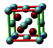
The N-V center defect consists of a nitrogen atom in place of a carbon atom next to a vacancy (empty space instead of an atom) within the diamond’s lattice structure.[16] Recent advances (up to 2019) in the field of nanodiamonds in quantum sensing applications using NVs have been summarized in the following review.[17]
Applying a microwave pulse to such a defect switches the direction of its electron spin. Applying a series of such pulses (Walsh decoupling sequences) causes them to act as filters. Varying the number of pulses in a series switched the spin direction a different number of times.[16] They efficiently extract spectral coefficients while suppressing decoherence, thus improving sensitivity.[18] Signal-processing techniques were used to reconstruct the entire magnetic field.[16]
The prototype used a 3 mm-diameter square diamond, but the technique can scale down to tens of nanometers.[16]
Nano-abrasive edit
Nanodiamonds share the hardness and chemical stability of visible-scale diamonds, making them candidates for applications such as polishes and engine oil additives for improved lubrication.[3]
Medical edit
Diamond nanoparticles have the potential to be used in myriad biological applications and due to their unique properties such as inertness and hardness, nanodiamonds may prove to be a better alternative to the traditional nanomaterials currently utilized to carry drugs, coat implantable materials, and synthesize biosensors and biomedical robots.[19] The low cytotoxicity of diamond nanoparticles affirms their utilization as biologically compatible materials.[19]
In vitro studies exploring the dispersion of diamond nanoparticles in cells have revealed that most diamond nanoparticles exhibit fluorescence and are uniformly distributed.[20] Fluorescent nanodiamond particles can be mass produced through irradiating diamond nanocrystallites with helium ions.[21] Fluorescent nanodiamond is photostable, chemically inert, and has extended fluorescent lifetime, making it a great candidate for many biological applications.[22] Studies have shown that small photoluminescent diamond nanoparticles that remain free in the cytosol are excellent contenders for the transport of biomolecules.[23]
In-vitro diagnostics edit
Nanodiamonds containing nitrogen-vacancy defects have been used as an ultrasensitive label for in vitro diagnostics, using a microwave field to modulate emission intensity and frequency-domain analysis to separate the signal from background autofluorescence.[24] Combined with recombinase polymerase amplification, nanodiamonds enable single-copy detection of HIV-1 RNA on a low-cost lateral flow test format.
Drug delivery edit
Diamond nanoparticles of ~5 nm in size offer a large accessible surface and tailorable surface chemistry. They have unique optical, mechanical and thermal properties and are non-toxic. The potential of nanodiamond in drug delivery has been demonstrated, fundamental mechanisms, thermodynamics and kinetics of drug adsorption on nanodiamond are poorly understood. Important factors include purity, surface chemistry, dispersion quality, temperature and ionic composition.
Nanodiamonds (with attached molecules) are able to penetrate the blood–brain barrier that isolates the brain from most insults. In 2013 doxorubicin molecules (a popular cancer-killing drug) were bonded to nanodiamond surfaces, creating the drug ND-DOX. Tests showed that tumors were unable to eject the compound, increasing the drug's ability to impact the tumor and reducing side-effects.[3]
Larger nanodiamonds, due to their "high uptake efficiency", have the potential to serve as cellular labels.[23] Studies have concluded that diamond nanoparticles are similar to carbon nanotubes and upon being treated with surfactants, the stability and biocompatibility of both carbon nanotubes and the nanodiamonds in solution greatly increase.[20] In addition, the ability to surface functionalize nanodiamonds of small diameters provides various possibilities for diamond nanoparticles to be utilized as biolabels with potentially low cytotoxicity.[20]
Catalysis edit
Decreasing particle size and functionalizing their surfaces[20] may allow such surface-modified diamond nanoparticles to deliver proteins, which can then provide an alternative to traditional catalysts.[25]
Skin care edit
Nanodiamonds are well-absorbed by human skin. They also absorb more of the ingredients in skin care products than skin itself. Thus they cause more of the ingredients to penetrate the deeper layers of the skin. Nanodiamonds also form strong bonds with water, helping to hydrate the skin.[3]
Surgery edit
During jaw and tooth repair operations, doctors normally use invasive surgery to stick a sponge containing bone-growth-stimulating proteins near the affected area. However, nanodiamonds bind to both bone morphogenetic protein and fibroblast growth factor, both of which encourage bone and cartilage to rebuild and can be delivered orally.[3] Nanodiamond has also been successfully incorporated into gutta percha in root canal therapy.[26]
Blood testing edit
Defected nanodiamonds can measure the orientation of electron spins in external fields and thus measure their strength. They can electrostatically absorb ferritin proteins on the diamond surface where their numbers can be measured directly as well as the number of iron atoms (as many as 4,500) that make up the protein.[3]
Electronics and sensors edit
Sensor edit
Naturally occurring defects in nanodiamonds called nitrogen-vacancy (N-V) centers, have been used to measure changes over time in weak magnetic fields, much like a compass does with Earth's magnetic field. The sensors can be used at room temperature, and since they consist entirely of carbon, they could be injected into living cells without causing them any harm, Paola Cappellaro says.[16] Moreover, nanodiamond can be exploited as sensor for some specific analytes. Boron-doped diamond (BDD) produced by energy-assisted (plasma or hot filament, HF) Chemical Vapor Deposition (CVD) processes is a good candidatein Dopamine detection, however it is not selective towards some interferents. This issue, can be overcome via further post-synthesis treatments for BDD surface modifications including anodization, hydrogen plasma, etching into porous forms, carbon-based nanomaterials, polymer films and nanoparticles. Recent studies,[27] propose a new approach for the realization of Titanium doped diamond-based electrodes with a native selectivity towards dopamine, through substrate pre-treatments (lapping, electropolishing and chemical etching) instead of post-process treatments. Moreover, Nanodiamond has been proven to modify some electronic properties of polymer-based matrix.[28] Those modifications, which can be summarised as an increase in the ionic conductivity of the system, thus of a decrease in the impedance, are likely due to the presence of functional groups on the nanodiamond particle surface. Those groups can interact with polymer chains, thus facilitating ionic exchanges.
Nanomechanical sensor and nanoelectromechanical system (NEMS) edit
Recent studies have shown that nanoscale diamonds can be bent to a local maximum tensile elastic strain in excess of 9%,[29] with the corresponding maximum tensile stress reached ~100 gigapascals, making them ideal for high-performance nanomechanical sensor and NEMS applications.
Optical computing edit
Nanodiamonds offer an alternative to photonic metamaterials for optical computing. The same single-defect nanodiamonds that can be used to sense magnetic fields can also use combinations of green and infrared light to enable/disrupt light transmission, allowing the construction of transistors and other logic elements.[3]
Quantum computing edit
Nanodiamonds with NV centers may serve as a solid-state alternative to trapped ions for room-temperature quantum computing.[3]
Imaging edit
Fluorescent nanodiamonds offer a stable reference for the quality control purposes in fluorescence and multiharmonic imaging systems. [30]
Prizes and awards edit
- 2012 Ig Nobel Peace Prize: The SKN Company, for converting old Russian ammunition into new diamonds
- In 2015 Amanda Barnard, Science Leader of Australia's Office of the Chief Executive (OCE), The Commonwealth Scientific and Industrial Research Organisation (CSIRO), received the Theory Prize at the Foresight Institutes' Feynman Awards for nanotechnology. Using theoretical and computational methods, Amanda Barnard increased understanding of the structure and stability of carbon nanostructures and the role that shape plays in establishing properties and interactions under different conditions. The Prize announced focused on her work on diamond nanoparticles (nanodiamonds).[31]
See also edit
- Aggregated diamond nanorod, a nanocrystalline form of diamond also known as nanodiamond or hyperdiamond
- Detonation nanodiamond
- Fiveling, twinned structure common in nanodiamonds
- Icosahedral twins, structure common in nanodiamonds
References edit
- ^ a b c Ohfuji, Hiroaki; Irifune, Tetsuo; Litasov, Konstantin D.; Yamashita, Tomoharu; Isobe, Futoshi; Afanasiev, Valentin P.; Pokhilenko, Nikolai P. (2015). "Natural occurrence of pure nano-polycrystalline diamond from impact crater". Scientific Reports. 5: 14702. Bibcode:2015NatSR...514702O. doi:10.1038/srep14702. PMC 4589680. PMID 26424384.
- ^ Chung, P.-H.; Perevedentseva, E.; Cheng, C.-L. (2007). "The particle size-dependent photoluminescence of nanodiamonds". Surface Science. 601 (18): 3866–3870. Bibcode:2007SurSc.601.3866C. doi:10.1016/j.susc.2007.04.150.
- ^ a b c d e f g h i Feinberg, Ashley (April 9, 2014). "How These Microscopic Diamonds Are Going to Shape the Future". Gizmodo.
- ^ Mochalin, V. N.; Shenderova, O.; Ho, D.; Gogotsi, Y. (2011). "The properties and applications of nanodiamonds". Nature Nanotechnology. 7 (1): 11–23. doi:10.1038/nnano.2011.209. PMID 22179567.
- ^ Danilenko, V. V. (2004). "On the history of the discovery of nanodiamond synthesis". Physics of the Solid State. 46 (4): 595–599. Bibcode:2004PhSS...46..595D. doi:10.1134/1.1711431. S2CID 121038737.
- ^ Zou, Q.; Li, Y.G.; Zou, L.H.; Wang, M.Z. (2009). "Characterization of structures and surface states of the nanodiamond synthesized by detonation". Materials Characterization. 60 (11): 1257–1262. doi:10.1016/j.matchar.2009.05.008.
- ^ Paci, Jeffrey T.; Man, Han B.; Saha, Biswajit; Ho, Dean; Schatz, George C. (2013). "Understanding the Surfaces of Nanodiamonds". The Journal of Physical Chemistry C. 117 (33): 17256–17267. doi:10.1021/jp404311a.
- ^ Fang, Xiaowen; Mao, Jingdong; Levin, E. M.; Schmidt-Rohr, Klaus (2009). "Nonaromatic Core−Shell Structure of Nanodiamond from Solid-State NMR Spectroscopy". Journal of the American Chemical Society. 131 (4): 1426–1435. doi:10.1021/ja8054063. PMID 19133766.
- ^ Rondin, L.; Dantelle, G.; Slablab, A.; Grosshans, F.; Treussart, F.; Bergonzo, P.; Perruchas, S.; Gacoin, T.; Chaigneau, M.; Chang, H.-C.; Jacques, V.; Roch, J.-F. (2010). "Surface-induced charge state conversion of nitrogen-vacancy defects in nanodiamonds". Physical Review B. 82 (11): 115449. arXiv:1008.2276. Bibcode:2010PhRvB..82k5449R. doi:10.1103/PhysRevB.82.115449. S2CID 119217590.
- ^ "Ultrasonic Synthesis of Nanodiamonds". www.hielscher.com.
- ^ Kharisov, Boris I.; Kharissova, Oxana V.; Chávez-Guerrero, Leonardo (2010). "Synthesis Techniques, Properties, and Applications of Nanodiamonds". Synthesis and Reactivity in Inorganic, Metal-Organic, and Nano-Metal Chemistry. 40: 84–101. doi:10.3109/10799890903555665 (inactive 31 January 2024).
{{cite journal}}: CS1 maint: DOI inactive as of January 2024 (link) - ^ Fang, Leiming; Ohfuji, Hiroaki; Irifune, Tetsuo (2013). "A Novel Technique for the Synthesis of Nanodiamond Powder". Journal of Nanomaterials. 2013: 1–4. doi:10.1155/2013/201845. S2CID 54026958.
- ^ a b c d Holt, Katherine B. (2007). "Diamond at the nanoscale: Applications of diamond nanoparticles from cellular biomarkers to quantum computing". Philosophical Transactions of the Royal Society A: Mathematical, Physical and Engineering Sciences. 365 (1861): 2845–2861. Bibcode:2007RSPTA.365.2845H. doi:10.1098/rsta.2007.0005. PMID 17855222. S2CID 8185618.
- ^ Kumar, Ajay; Ann Lin, Pin; Xue, Albert; Hao, Boyi; Khin Yap, Yoke; Sankaran, R. Mohan (21 October 2013). "Formation of nanodiamonds at near-ambient conditions via microplasma dissociation of ethanol vapour". Nature Communications. 4 (1): 2618. Bibcode:2013NatCo...4.2618K. doi:10.1038/ncomms3618. PMID 24141249. S2CID 26552314.
- ^ Nee, Chen-Hon; Yap, Seong-Ling; Tou, Teck-Yong; Chang, Huan-Cheng; Yap, Seong-Shan (23 September 2016). "Direct synthesis of nanodiamonds by femtosecond laser irradiation of ethanol". Scientific Reports. 6 (1): 33966. Bibcode:2016NatSR...633966N. doi:10.1038/srep33966. PMC 5034281. PMID 27659184.
- ^ a b c d e "Using nanodiamonds to precisely detect neural signals". KurzweilAI. January 27, 2014.
- ^ Radtke, Mariusz; Bernardi, Ettore; Slablab, Abdallah; Nelz, Richard; Neu, Elke (9 September 2019). "Nanoscale sensing based on nitrogen vacancy centersin single crystal diamond and nanodiamonds:achievements and challenges". arXiv:1909.03719v1 [physics.app-ph].
- ^ Cooper, A.; Magesan, E.; Yum, H. N.; Cappellaro, P. (2014). "Time-resolved magnetic sensing with electronic spins in diamond". Nature Communications. 5: 3141. arXiv:1305.6082. Bibcode:2014NatCo...5.3141C. doi:10.1038/ncomms4141. PMID 24457937. S2CID 14914691.
- ^ a b Schrand, Amanda M.; Huang, Houjin; Carlson, Cataleya; Schlager, John J.; Ōsawa, Eiji; Hussain, Saber M.; Dai, Liming (2007). "Are Diamond Nanoparticles Cytotoxic?". The Journal of Physical Chemistry B. 111 (1): 2–7. doi:10.1021/jp066387v. PMID 17201422.
- ^ a b c d Neugart, Felix; Zappe, Andrea; Jelezko, Fedor; Tietz, C.; Boudou, Jean Paul; Krueger, Anke; Wrachtrup, Jörg (2007). "Dynamics of Diamond Nanoparticles in Solution and Cells". Nano Letters. 7 (12): 3588–3591. Bibcode:2007NanoL...7.3588N. doi:10.1021/nl0716303. PMID 17975943.
- ^ Chang, Yi-Ren; Lee, Hsu-Yang; Chen, Kowa; Chang, Chun-Chieh; Tsai, Dung-Sheng; Fu, Chi-Cheng; Lim, Tsong-Shin; Fang, Chia-Yi; Han, Chau-Chung; Chang, Huan-Cheng; Fann, Wunshain (2008). "Mass Production and Dynamic Imaging of Fluorescent Nanodiamonds". Nature Nanotechnology. 3 (5): 284–288. doi:10.1038/nnano.2008.99. PMID 18654525.
- ^ Yu, Shu-Jung; Kang, Ming-Wei; Chang, Huan-Cheng; Chen, Kuan-Ming; Yu, Yueh-Chung (2005). "Bright Fluorescent Nanodiamonds: No Photobleaching and Low Cytotoxicity". Journal of the American Chemical Society. 127 (50): 17604–17605. doi:10.1021/ja0567081. PMID 16351080.
- ^ a b Faklaris, O.; Joshi, V.; Irinopoulou, T.; Tauc, P.; Sennour, M.; Girard, H.; Gesset, C.; Arnault, J. C.; Thorel, A.; Boudou, J. P.; Curmi, P. A.; Treussart, F. (2009). "Photoluminescent diamond nanoparticles for cell labeling: Study of the uptake mechanism in mammalian cells". ACS Nano. 3 (12): 3955–62. arXiv:0907.1148. doi:10.1021/nn901014j. PMID 19863087. S2CID 1261084.
- ^ Miller, Benjamin S.; Bezinge, Léonard; Gliddon, Harriet D.; Huang, Da; Dold, Gavin; Gray, Eleanor R.; Heaney, Judith; Dobson, Peter J.; Nastouli, Eleni; Morton, John J. L.; McKendry, Rachel A. (2020). "Spin-enhanced nanodiamond biosensing for ultrasensitive diagnostics". Nature. 587 (7835): 588–593. Bibcode:2020Natur.587..588M. doi:10.1038/s41586-020-2917-1. PMID 33239800. S2CID 227176732.
- ^ Kossovsky, Nir; Gelman, Andrew; Hnatyszyn, H. James; Rajguru, Samir; Garrell, Robin L.; Torbati, Shabnam; Freitas, Siobhan S. F.; Chow, Gan-Moog (1995). "Surface-Modified Diamond Nanoparticles as Antigen Delivery Vehicles". Bioconjugate Chemistry. 6 (5): 507–511. doi:10.1021/bc00035a001. PMID 8974446.
- ^ Lee, Dong-Keun; Lee, Theordore; Liang, Zhangrui; Hsiou, Desiree; Miya, Darron; Wu, Brian; Osawa, Eiji; Chow, Edward Kai-Hua; Sung, Eric C; Kang, Mo K.; Ho, Dean (2017). "Clinical validation of a nanodiamond-embedded thermoplastic biomaterial". PNAS. 114 (45): E9445–E9454. Bibcode:2017PNAS..114E9445L. doi:10.1073/pnas.1711924114. PMC 5692571. PMID 29078364.
- ^ Tamburri, Carcione (2023). "Pretreatment strategies of titanium substrates to modulate the electrochemical properties of CVD-grown Ti-doped diamond electrodes for dopamine detection". Surface and Coatings Technology. 467 (129662). doi:10.1016/j.surfcoat.2023.129662.
- ^ Orlanducci, Palmieri (2022). "A Sustainable Hydroxypropyl Cellulose-Nanodiamond Composite for Flexible Electronic Applications". Gels. 12 (8): 783. doi:10.3390/gels8120783. PMC 9777684. PMID 36547307.
- ^ Banerjee, Amit; et al. (2018). "Ultralarge elastic deformation of nanoscale diamond". Science. 360 (6386): 300–302. Bibcode:2018Sci...360..300B. doi:10.1126/science.aar4165. PMID 29674589. S2CID 5047604.
- ^ Žurauskas, Mantas; Alex, Aneesh; Park, Jaena; Hood, Steve R.; Boppart, Stephen A. (2021-11-03). "Fluorescent nanodiamonds for characterization of nonlinear microscopy systems". Photonics Research. 9 (12). Optica: 2309–2318. doi:10.1364/prj.434236. ISSN 2327-9125. PMC 10174270. PMID 37181134. S2CID 239280518.
- ^ "2014 Foresight Institute Feynman Prize". Foresight Institute. April 2015.