


Summary
Optical tomography is a form of computed tomography that creates a digital volumetric model of an object by reconstructing images made from light transmitted and scattered through an object.[1] Optical tomography is used mostly in medical imaging research. Optical tomography in industry is used as a sensor of thickness and internal structure of semiconductors.[2]
| Optical tomography | |
|---|---|
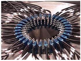 A fiber-optic array for breast cancer detection by way of Diffuse Optical Tomography. | |
| MeSH | D041622 |
Principle edit
Optical tomography relies on the object under study being at least partially light-transmitting or translucent, so it works best on soft tissue, such as breast and brain tissue.
The high scatter-based attenuation involved is generally dealt with by using intense, often pulsed or intensity modulated, light sources, and highly sensitive light sensors, and the use of infrared light at frequencies where body tissues are most transmissive. Soft tissues are highly scattering but weakly absorbing in the near-infrared and red parts of the spectrum, so that this is the wavelength range usually used.
Types edit
Diffuse optical tomography edit
In near-infrared diffuse optical tomography (DOT), transmitted diffuse photons are collected and a diffusion equation is used to reconstruct an image from them.[3]
Time-of-flight diffuse optical tomography edit
A variant of optical tomography uses optical time-of-flight sampling as an attempt to distinguish transmitted light from scattered light.[4] This concept has been used in several academic and commercial systems for breast cancer imaging and cerebral measurement. The key to separation of absorption from scatter is the use of either time-resolved or frequency domain data which is then matched with a diffusion theory based estimate of how the light propagated through the tissue. The measurement of time of flight or frequency domain phase shift is essential to allow separation of absorption from scatter with reasonable accuracy.[citation needed]
Fluorescence molecular tomography edit
In fluorescence molecular tomography, the fluorescence signal transmitted through the tissue is normalized by the excitation signal transmitted through the tissue, and so many of the fluorescence tomography systems do not require the use of time-resolved or frequency domain data, although research is still ongoing in this area. Since the applications of fluorescent molecules in humans are fairly limited, most of the work in fluorescence tomography has been in the realm of pre-clinical cancer research. Both commercial systems and academic research have been shown to be effective in tracking tumor protein expression and production, and tracking response to therapies.[citation needed]
Confocal diffuse tomography edit
Confocal diffuse tomography uses a powerful laser to illuminate a sample through a scattering medium, followed by deconvolution with a calibrated diffusion operator to estimate a volume without the effects of diffusive scattering and subsequent application of a confocal inverse filter to recover the sample image.[5][6]
See also edit
References edit
- ^ Optical+Tomography at the U.S. National Library of Medicine Medical Subject Headings (MeSH)
- ^ ^ Wojtek J. Walecki and Fanny Szondy, "Integrated quantum efficiency, reflectance, topography and stress metrology for solar cell manufacturing", Sunrise Optical LLC, Proc. SPIE 7064, 70640A (2008); doi:10.1117/12.797541
- ^ Hoshi, Yoko; Yamada, Yukio (2016-07-13). "Overview of diffuse optical tomography and its clinical applications". Journal of Biomedical Optics. 21 (9): 091312. Bibcode:2016JBO....21i1312H. doi:10.1117/1.JBO.21.9.091312. ISSN 1083-3668. PMID 27420810.
- ^ Lyons, Ashley; Tonolini, Francesco; Boccolini, Alessandro; Repetti, Audrey; Henderson, Robert; Wiaux, Yves; Faccio, Daniele (August 2019). "Computational time-of-flight diffuse optical tomography". Nature Photonics. 13 (8): 575–579. arXiv:1808.01135. Bibcode:2019NaPho..13..575L. doi:10.1038/s41566-019-0439-x. ISSN 1749-4885. S2CID 118707188.
- ^ Lindell, David B.; Wetzstein, Gordon (December 2020). "Three-dimensional imaging through scattering media based on confocal diffuse tomography". Nature Communications. 11 (1): 4517. Bibcode:2020NatCo..11.4517L. doi:10.1038/s41467-020-18346-3. ISSN 2041-1723. PMC 7481188. PMID 32908155.
- ^ "Confocal Diffuse Tomography | Nature Communications 2020 - YouTube". www.youtube.com. Retrieved 2021-02-10.
Further reading edit
External links edit
- Optical tomography at Imperial College, London
- Optical tomography at University College, London
- "Boobs, Babes and Blood" - Article on physics.org