


Summary
The palatovaginal canal (also pharyngeal canal) is a small canal formed between the sphenoidal process of palatine bone, and vaginal process of sphenoid bone.[1]: 508 It connects the pterygopalatine fossa and[1]: 370 and nasal cavity.[citation needed] It transmits the pharyngeal nerve (pharyngeal branch of maxillary nerve), and the pharyngeal branch of maxillary artery.[1]: 508
| Palatovaginal canal | |
|---|---|
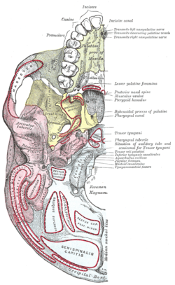 Base of skull. Inferior surface. (Pharyngeal canal labeled at right, tenth label from the top.) | |
| Details | |
| Identifiers | |
| Latin | c. pharyngeus, c. palatovaginalis |
| TA98 | A02.1.00.064 |
| TA2 | 466 |
| FMA | 54372 |
| Anatomical terminology [edit on Wikidata] | |
Anatomy edit
Its proximal opening is situated inferoposteriorly in the pterygopalatine fossa.[citation needed]
Its distal opening is situated in the nasal cavity at the root of the pterygoid process near the lateral margin of the ala of vomer.[citation needed]
Variation edit
An inconstant vomerovaginal canal may lie between the ala of the vomer and the vaginal process of the sphenoid bone, medial to the palatovaginal canal, and lead into the anterior end of the palatovaginal canal.[citation needed]
Contents edit
The pharyngeal branch of the maxillary artery supplies the nasopharynx,[citation needed] posterior part of the roof of the nasal cavity, sphenoid sinus, and pharyngotympanic tube.[2]
References edit
![]() This article incorporates text in the public domain from page 180 of the 20th edition of Gray's Anatomy (1918)
This article incorporates text in the public domain from page 180 of the 20th edition of Gray's Anatomy (1918)
External links edit
- Rumboldt Z, Castillo M, Smith JK (2002). "The palatovaginal canal: can it be identified on routine CT and MR imaging?". AJR Am J Roentgenol. 179 (1): 267–72. doi:10.2214/ajr.179.1.1790267. PMID 12076948.