


Summary
The pterygoid hamulus is a hook-like process at the lower extremity of the medial pterygoid plate of the sphenoid bone of the skull. It is the superior origin of the pterygomandibular raphe, and the levator veli palatini muscle.
| Pterygoid hamulus | |
|---|---|
 Sphenoid bone. Anterior and inferior surfaces. (Hamulus labeled at bottom left.) | |
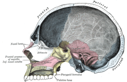 Sagittal section of skull. (Sphenoid is in yellow, and pterygoid hamulus labeled at bottom center.) | |
| Details | |
| Part of | sphenoid bone of skull |
| System | skeletal |
| Identifiers | |
| Latin | hamulus pterygoideus |
| TA98 | A02.1.05.051 |
| TA2 | 637 |
| FMA | 54722 |
| Anatomical terms of bone [edit on Wikidata] | |
Structure edit
The pterygoid hamulus is part of the medial pterygoid plate of the sphenoid bone of the skull. Its tip is rounded off.[1] It has an average length of 7.2 mm, an average depth of 1.4 mm, and an average width of 2.3 mm.[1] The tendon of tensor veli palatini muscle glides around it.[1]
Function edit
The pterygoid hamulus is the superior origin of the pterygomandibular raphe. It is also the origin of levator veli palatini muscle.[1]
Clinical significance edit
Rarely, the pterygoid hamulus may be enlarged, which may cause mouth pain.[2]
See also edit
References edit
![]() This article incorporates text in the public domain from page 151 of the 20th edition of Gray's Anatomy (1918)
This article incorporates text in the public domain from page 151 of the 20th edition of Gray's Anatomy (1918)
- ^ a b c d Putz, R.; Kroyer, A. (1 January 1999). "Functional morphology of the pterygoid hamulus". Anatomischer Anzeiger. 181 (1): 85–88. doi:10.1016/s0940-9602(99)80099-5. ISSN 1618-0402. PMID 10081567.
- ^ Sasaki, T.; Imai, Y.; Fujibayashi, T. (2001). "A case of elongated pterygoid hamulus syndrome". Oral Diseases. 7 (2): 131–133. doi:10.1034/j.1601-0825.2001.70212.x. ISSN 1601-0825. PMID 11355439.
External links edit
- Anatomy figure: 22:4b-05 at Human Anatomy Online, SUNY Downstate Medical Center
- "Anatomy diagram: 05287.011-1". Roche Lexicon - illustrated navigator. Elsevier. Archived from the original on 2013-04-22.