


Summary
Respiratory bronchiolitis is a lung disease associated with tobacco smoking.[1] In pathology, it is defined by the presence of "smoker's macrophages".[1] When manifesting significant clinical symptoms it is referred to as respiratory bronchiolitis interstitial lung disease (RB-ILD).[1]
| Respiratory bronchiolitis | |
|---|---|
| Other names | RB-ILD |
 | |
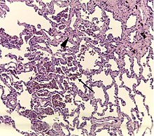
| A "smoker's macrophage", with yellow to light brown and finely granular cytoplasmic pigment. | |
| Specialty | Pulmonology |
Diagnosis edit
Diagnosis of respiratory bronchiolitis requires a correlation of clinical, radiologic and pathologic findings:[1]
- Clinical: Symptoms and pulmonary function testing
- Radiologic: Chest radiograph and high-resolution computed tomography
- Pathologic: Lung biopsy with "smoker's macrophages" limited to distal airspaces and peribronchiolar airspaces, and minimal to absent peribronchiolar interstitial fibrotic thickening
Respiratory bronchiolitis interstitial lung disease edit

Respiratory bronchiolitis interstitial lung disease is respiratory bronchiolitis that manifests as a clinically significant interstitial lung disease.[1] It is a form of idiopathic interstitial pneumonia associated with smoking.[3]
It is a histological finding, not a pathological description. When associated with disease, it is known as "Respiratory bronchiolitis-associated interstitial lung disease" or "RB-ILD".[4] Also, this disease is predominantly found in the upper lobe with centrilobar ground glass nodules. Importantly, no fibrosis is involved, just bronchial wall thickening. Treatment is to stop smoking.
The appearance is similar to desquamative interstitial pneumonia, and some have suggested that the two conditions are caused by the same processes.[5]
See also edit
References edit
- ^ a b c d e William Perry, M.D., M.P.H., Kristine Konopka, M.D. "Respiratory bronchiolitis". Pathology Outlines.
{{cite web}}: CS1 maint: multiple names: authors list (link) Topic Completed: 1 July 2020. Minor changes: 1 July 2020 - ^ Sousa, Célia; Rodrigues, Márcio; Carvalho, André; Viamonte, Bárbara; Cunha, Rui; Guimarães, Susana; de Moura, Conceição Souto; Morais, António; Pereira, José Miguel (2019). "Diffuse smoking-related lung diseases: insights from a radiologic-pathologic correlation". Insights into Imaging. 10 (1): 73. doi:10.1186/s13244-019-0765-z. ISSN 1869-4101. PMC 6635572. PMID 31312909.
- This article is distributed under the terms of the Creative Commons Attribution 4.0 International License (http://creativecommons.org/licenses/by/4.0/) - ^ "Idiopathic Interstitial Pneumonias: Interstitial Lung Diseases: Merck Manual Professional". Retrieved 2008-12-09.
- ^ Cotran, Ramzi S.; Kumar, Vinay; Fausto, Nelson; Nelso Fausto; Robbins, Stanley L.; Abbas, Abul K. (2005). Robbins and Cotran pathologic basis of disease. St. Louis, Mo: Elsevier Saunders. p. 741. ISBN 0-7216-0187-1.
- ^ Heyneman LE, Ward S, Lynch DA, Remy-Jardin M, Johkoh T, Müller NL (December 1999). "Respiratory bronchiolitis, respiratory bronchiolitis-associated interstitial lung disease, and desquamative interstitial pneumonia: different entities or part of the spectrum of the same disease process?". AJR Am J Roentgenol. 173 (6): 1617–22. doi:10.2214/ajr.173.6.10584810. PMID 10584810.