


Summary
The sacrospinous ligament (small or anterior sacrosciatic ligament) is a thin, triangular ligament in the human pelvis. The base of the ligament is attached to the outer edge of the sacrum and coccyx, and the tip of the ligament attaches to the spine of the ischium, a bony protuberance on the human pelvis. Its fibres are intermingled with the sacrotuberous ligament.
| Sacrospinous ligament | |
|---|---|
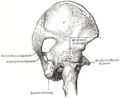
 Articulations of pelvis, anterior view, with greater sciatic foramen (labeled in red) and its boundaries. | |
| Details | |
| From | Ischial spine |
| To | Sacrum |
| Identifiers | |
| Latin | ligamentum sacrospinale |
| TA98 | A03.6.03.007 |
| TA2 | 1852 |
| FMA | 21485 |
| Anatomical terminology [edit on Wikidata] | |
Structure edit
The sacrotuberous ligament passes behind the sacrospinous ligament. In its entire length, the sacrospinous ligament covers the equally triangular coccygeus muscle, to which its closely connected.[1]
Function edit
The presence of the ligament in the greater sciatic notch creates an opening (foramen), the greater sciatic foramen, and also converts the lesser sciatic notch into the lesser sciatic foramen.[2] The greater sciatic foramen lies above the ligament, and the lesser sciatic foramen lies below it.
The pudendal vessels and nerve pass behind the sacrospinous ligament directly medially and inferiorly to the ischial spine. The inferior gluteal artery, from a branch of the internal iliac artery, pass behind the sciatic nerve and the sacrospinous ligament and is left uncovered in a small opening above the top of the sacrospinous ligament. The coccygeal branch of the inferior gluteal artery passes behind the mid-portion of the sacrospinous ligament and pierces the sacrotuberous ligament at multiple locations. The main body of the inferior gluteal artery leaves the pelvis posteriorly to the upper border of the sacrospinous ligament, to follow the inferior portion of the sciatic nerve out of the greater sciatic foramen.[3]
The main function of the ligament is to prevent rotation of the ilium past the sacrum. Laxity of this ligament and the sacrotuberous ligament allows this rotation to occur. Stresses to these ligaments occur most often when leaning forward or getting out of a chair.[citation needed]
Clinical significance edit
Vaginal prolapse or uterine prolapse may occur in women when other pelvic ligaments and supportive structures are weakened. One treatment is sacrospinous fixation. In this surgery, the apex of the vagina is sutured to the sacrospinous ligament, which may offer a sturdier support than weakened pelvic ligaments, ideally preventing further prolapse.[4]
Additional images edit
-
 Nélaton's line and Bryant's triangle.
 -
 Articulations of pelvis. Posterior view.


Notes edit
References edit
![]() This article incorporates text in the public domain from page 309 of the 20th edition of Gray's Anatomy (1918)
This article incorporates text in the public domain from page 309 of the 20th edition of Gray's Anatomy (1918)
- Thompson, Jason R.; Gibb, John S.; Genadry, Rene; Burrows, Lara; Lambrou, Nicholas; Buller, Jerome L. (1999). "Anatomy of Pelvic Arteries Adjacent to the Sacrospinous Ligament: Importance of the Coccygeal Branch of the Inferior Gluteal Artery". Obstetrics & Gynecology. 94 (6). Baltimore, Maryland.: Johns Hopkins University: 973–977. doi:10.1016/s0029-7844(99)00418-4. PMID 10576185.
- Platzer, Werner (2004). Color Atlas of Human Anatomy, Vol 1: Locomotor system (5th ed.). Thieme. ISBN 3-13-533305-1. (ISBN for the Americas 1-58890-159-9.)
- Vasavada, Sandip P.; Appell, Rodney; Sand, Peter K.; Raz, Shlomo (2004). Female Urology, Urogynecology, and Voiding Dysfunction. Informa Health Care. ISBN 0-8247-5426-3.
External links edit
- Anatomy figure: 17:02-03 at Human Anatomy Online, SUNY Downstate Medical Center - "Posterior view of the bones and ligaments of the hip joint."
- Johns Hopkins