


Summary
Stereocilia (or stereovilli or villi) are non-motile apical cell modifications. They are distinct from cilia and microvilli, but are closely related to microvilli. They form single "finger-like" projections that may be branched, with normal cell membrane characteristics. They contain actin. Stereocilia are found in the vas deferens, the epididymis, and the sensory cells of the inner ear.
| Stereocilia | |
|---|---|
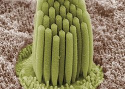 Stereocilia of frog inner ear | |
| Identifiers | |
| MeSH | D059547 |
| TH | H1.00.01.1.01013 |
| Anatomical terms of microanatomy [edit on Wikidata] | |
Structure edit
Stereocilia are cylindrical and non-motile. They are much longer and thicker than microvilli, form single "finger-like" projections that may be branched, and have more of the characteristics of the cellular membrane proper. Like microvilli, they contain actin[1] and lack an axoneme. This distinguishes them from cilia.
They do not have a basal body at their base since they do not contain microtubules. They may or may not be covered by a glycocalyx coating. They have no fixed arrangement, different to the structure present in kinocilium.
Organs containing stereocilia edit
Stereocilia are found in:
- the vas deferens.
- the epididymis. Some consider epididymal stereocilia to be a variant of microvilli, rather than their own distinct type of structure.[2]
- the sensory (hair) cells of the inner ear.
References edit
- ^ Tilney, Lewis G.; Tilney, Mary S.; DeRosier, David J. (November 1992). "Actin Filaments, Stereocilia, and Hair Cells: How Cells Count and Measure". Annual Review of Cell Biology. 8 (1): 257–274. doi:10.1146/annurev.cb.08.110192.001353. ISSN 0743-4634. PMID 1476800.
- ^ Krause J. William (July 2005). Krause's Essential Human Histology for Medical Students. Universal-Publishers. p. 37. ISBN 978-1-58112-468-2.
External links edit
- J.A. Illingworth, "Sensory Transducers" (Lectures, 2011), University of Leeds