


Summary
The sternoclavicular joint or sternoclavicular articulation is a synovial saddle joint between the manubrium of the sternum, and the clavicle, and the first costal cartilage. The joint possesses a joint capsule, and an articular disc, and is reinforced by multiple ligaments.[1]
| Sternoclavicular joint | |
|---|---|
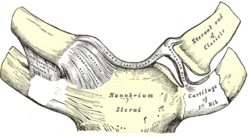 Sternoclavicular joint. Anterior view. | |
 Sternoclavicular joint visible near center but not labeled. | |
| Details | |
| Identifiers | |
| Latin | articulatio sternoclavicularis |
| MeSH | D013247 |
| TA98 | A03.5.04.001 |
| TA2 | 1750 |
| FMA | 25883 |
| Anatomical terminology [edit on Wikidata] | |
Structure edit
The joint is structurally classified as a synovial saddle joint and functionally classed as a diarthrosis and multiaxial joint. It is composed of two portions separated by an articular disc of fibrocartilage.[1]
The joint is formed by the sternal end of the clavicle, the clavicular notch of the sternum, and (the superior surface of) the costal cartilage of the first rib.[1] The articular surface of the clavicle is larger than that of the sternum, and is invested with a layer of cartilage, which is considerably thicker than that of the sternum.[1]
The joint receives arterial supply via branches of the internal thoracic artery and the suprascapular artery. It is innervated via the medial supraclavicular nerve (superficially), and the nerve to subclavius (deeply).[1]
Joint capsule edit
The joint capsule is thickened anteriorly and posteriorly, but is thinner superiorly and (especially) inferiorly, where it consists mostly of loose areolar connective tissue.[1]
Articular disc edit
The joint features a fibrocartilaginous articular disc which completely divides the joint to form two articular compartments.[1] the disc acts to increase the range of movement of the joint.[2]
Ligaments edit
The joint is reinforced by two intrinsic and two extrinsic ligaments.[1] The costoclavicular ligament is the main limitation to movement, and therefore the main stabilizer of the joint.[citation needed]
- Anterior sternoclavicular ligament (intrinsic)[1]
- Posterior sternoclavicular ligament (intrinsic)[1]
- Costoclavicular ligament (extrinsic)[1]
- Interclavicular ligament (extrinsic)[1]
Function edit
The sternoclavicular joint allows movement of the clavicle in three planes, predominantly in the anteroposterior and vertical planes, although some rotation also occurs. A description of movement would be elevation and depression. Muscles do not directly act on this joint, although almost all actions of the shoulder girdle or the scapula will cause some motion at this articulation.[citation needed]
The unique double-hinged articular disk found at the junction of the clavicular head and manubrium allows for movement between the clavicle and the disk during elevation and depression of the scapula. This disk also allows motion between the sternum (manubrium) and itself during protraction and retraction of the scapula.[3]
Clinical significance edit
Dislocation edit
Sternoclavicular dislocation is rare,[2] but may result from direct trauma to the clavicle or indirect forces applied to the shoulder.[4] Posterior dislocations deserve special attention, as they have the potential to be life-threatening because of the risk of damage to vital structures in the mediastinum;[5] surgery can be used to fix such dislocations, as they are unlikely to heal by themselves.[6] A spontaneous partial dislocation may also sometimes occur.[citation needed]
Other edit
In SAPHO syndrome there may be arthropathy of the sternoclavicular joint.[citation needed]
Septic arthritis may rarely affect the sternoclavicular joint.[citation needed]
See also edit
- Acromioclavicular joint
- Shoulder
- Shoulder girdle (Pectoral girdle)
- Shoulder joint
References edit
- ^ a b c d e f g h i j k l Gray's anatomy : the anatomical basis of clinical practice. Susan Standring (Forty-second ed.). [New York]. 2021. ISBN 978-0-7020-7707-4. OCLC 1201341621.
{{cite book}}: CS1 maint: location missing publisher (link) CS1 maint: others (link) - ^ a b Cadogan, Mike (February 2010). "Sternoclavicular Joint Dislocations". Life in the Fast Lane. Retrieved June 5, 2011.
- ^ Lippert, Lynn. Clinical Kinesiology and Anatomy, 4th edition; pg.95-96.
- ^ Arend CF. Ultrasound of the Shoulder. Master Medical Books, 2013. Free section on sternoclavicular joint dislocation available at ShoulderUS.com
- ^ Jougon, Jacques B.; Lepront, Denis J.; Dromer, Claire E.H. (1996). "Posterior dislocation of the sternoclavicular joint leading to mediastinal compression". The Annals of Thoracic Surgery. 61 (2): 711–3. doi:10.1016/0003-4975(95)00745-8. PMID 8572795.
- ^ Terra, Bernardo Barcellos; Rodrigues, Leandro Marano; Pádua, David Victoria Hoffmann; Martins, Marcelo Giovanini; Teixeira, João Carlos de Medeiros; De Nadai, Anderson (2015-07-01). "Sternoclavicular dislocation: case report and surgical technique". Revista Brasileira de Ortopedia (English Edition). 50 (4): 472–477. doi:10.1016/j.rboe.2015.06.019. ISSN 2255-4971. PMC 4563050. PMID 26401506.
![]() This article incorporates text in the public domain from page 313 of the 20th edition of Gray's Anatomy (1918)
This article incorporates text in the public domain from page 313 of the 20th edition of Gray's Anatomy (1918)
External links edit
- Overview at ouhsc.edu
- Anatomy figure: 10:01-08 at Human Anatomy Online, SUNY Downstate Medical Center