


Summary
The temporal lobe is one of the four major lobes of the cerebral cortex in the brain of mammals. The temporal lobe is located beneath the lateral fissure on both cerebral hemispheres of the mammalian brain.[3]
| Temporal lobe | |
|---|---|
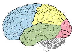
Lobes of the human brain (temporal lobe is shown in green) | |
 Section of brain showing upper surface of temporal lobe. | |
| Details | |
| Part of | Cerebrum |
| Artery | |
| Vein | |
| Identifiers | |
| Latin | lobus temporalis |
| MeSH | D013702 |
| NeuroNames | 125 |
| NeuroLex ID | birnlex_1160 |
| TA98 | A14.1.09.136 |
| TA2 | 5488 |
| FMA | 61825 |
| Anatomical terms of neuroanatomy [edit on Wikidata] | |
The temporal lobe is involved in processing sensory input into derived meanings for the appropriate retention of visual memory, language comprehension, and emotion association.[4]: 21 Temporal refers to the head's temples.
Structure edit
The temporal lobe consists of structures that are vital for declarative or long-term memory. Declarative (denotative) or explicit memory is conscious memory divided into semantic memory (facts) and episodic memory (events).[4]: 194 Medial temporal lobe structures that are critical for long-term memory include the hippocampus, along with the surrounding hippocampal region consisting of the perirhinal, parahippocampal, and entorhinal neocortical regions.[4]: 196 The hippocampus is critical for memory formation, and the surrounding medial temporal cortex is currently theorized to be critical for memory storage.[4]: 21 The prefrontal and visual cortices are also involved in explicit memory.[4]: 21
Research has shown that lesions in the hippocampus of monkeys results in limited impairment of function, whereas extensive lesions that include the hippocampus and the medial temporal cortex result in severe impairment.[5]
Function edit
Visual memories edit
The temporal lobe communicates with the hippocampus and plays a key role in the formation of explicit long-term memory modulated by the amygdala.[4]: 349
Processing sensory input edit
- Auditory
- Adjacent areas in the superior, posterior, and lateral parts of the temporal lobes are involved in high-level auditory processing. The temporal lobe is involved in primary auditory perception, such as hearing, and holds the primary auditory cortex.[6] The primary auditory cortex receives sensory information from the ears and secondary areas process the information into meaningful units such as speech and words.[6] The superior temporal gyrus includes an area (within the lateral fissure) where auditory signals from the cochlea first reach the cerebral cortex and are processed by the primary auditory cortex in the left temporal lobe.[citation needed]
- Visual
- The areas associated with vision in the temporal lobe interpret the meaning of visual stimuli and establish object recognition.[citation needed] The ventral part of the temporal cortices appears to be involved in high-level visual processing of complex stimuli such as faces (fusiform gyrus) and scenes (parahippocampal gyrus).[citation needed] Anterior parts of this ventral stream for visual processing are involved in object perception and recognition.[6]

Language recognition edit
The temporal lobe holds the primary auditory cortex, which is important for the processing of semantics in both language and vision in humans. Wernicke's area, which spans the region between temporal and parietal lobes, plays a key role (in tandem with Broca's area in the frontal lobe) in language comprehension,[7] whether spoken language or signed language. FMRI imaging shows these portions of the brain are activated by signed or spoken languages.[8][9] These areas of the brain are active in children's language acquisition[10] whether accessed via hearing a spoken language, watching a signed language, or via hand-over-hand tactile versions of a signed language[11]
The functions of the left temporal lobe are not limited to low-level perception but extend to comprehension, naming, and verbal memory.[12]
New memories edit
The medial temporal lobes (near the sagittal plane) are thought to be involved in encoding declarative long term memory.[4]: 194–199 The medial temporal lobes include the hippocampi, which are essential for memory storage, therefore damage to this area can result in impairment in new memory formation leading to permanent or temporary anterograde amnesia.[4]: 194–199
Clinical significance edit
Unilateral temporal lesion edit
- Contralateral homonymous upper quadrantanopia (sector anopsia)
- Complex hallucinations (smell, sound, vision, memory)
Dominant hemisphere edit
- Receptive aphasia
- Dyslexia
- Impaired verbal memory
- Word agnosia, word deafness
Non-dominant hemisphere edit
- Impaired non-verbal memory
- Impaired musical skills
Bitemporal lesions (additional features) edit
- Deafness
- Apathy (affective indifference)
- Impaired learning and memory
- Amnesia, Korsakoff syndrome, Klüver–Bucy syndrome
Damage edit
Individuals who suffer from medial temporal lobe damage have a difficult time recalling visual stimuli. This neurotransmission deficit is not due to lacking perception of visual stimuli, but rather to the inability to interpret what is perceived.[13] The most common symptom of inferior temporal lobe damage is visual agnosia, which involves impairment in the identification of familiar objects. Another less common type of inferior temporal lobe damage is prosopagnosia which is an impairment in the recognition of faces and distinction of unique individual facial features.[14]
Damage specifically to the anterior portion of the left temporal lobe can cause savant syndrome.[15]
Disorders edit
Pick's disease, also known as frontotemporal amnesia, is caused by atrophy of the frontotemporal lobe.[16] Emotional symptoms include mood changes, which the patient may be unaware of, including poor attention span and aggressive behavior towards themselves or others. Language symptoms include loss of speech, inability to read or write, loss of vocabulary and overall degeneration of motor ability.[17]
Temporal lobe epilepsy is a chronic neurological condition characterized by recurrent seizures; symptoms include a variety of sensory (visual, auditory, olfactory, and gustation) hallucinations, as well as an inability to process semantic and episodic memories.[18]
Schizophrenia is a severe psychotic disorder characterized by severe disorientation. Its most explicit symptom is the perception of external voices in the form of auditory hallucinations. The cause of such hallucinations has been attributed to deficits in the left temporal lobe, specifically within the primary auditory cortex.[19] Decreased gray matter, among other cellular deficits, contribute to spontaneous neural activity that affects the primary auditory cortex as if it were experiencing acoustic auditory input. The misrepresentation of speech in the auditory cortex results in the perception of external voices in the form of auditory hallucinations in schizophrenic patients.[20] Structural and functional MRI techniques have accounted for this neural activity by testing affected and non-affected individuals with external auditory stimuli.[19]
See also edit
References edit
- ^ a b c Starr, Philip A.; Barbaro, Nicholas M.; Larson, Paul S. (30 November 2008). Neurosurgical Operative Atlas: Functional Neurosurgery. Thieme. pp. 16, 26. ISBN 9781588903990.
- ^ Sekhar, Laligam N.; de Oliveira, Evandro (1999). Cranial Microsurgery: Approaches and Techniques. Thieme. p. 432. ISBN 9780865776982.
- ^ "Temporal Lobe". Langbrain. Rice University. Retrieved 2 January 2011.
- ^ a b c d e f g h Smith; Kosslyn (2007). Cognitive Psychology: Mind and Brain. New Jersey: Prentice Hall. pp. 21, 194–199, 349.
- ^ Squire, LR; Stark, CE; Clark, RE (2004). "The medial temporal lobe" (PDF). Annual Review of Neuroscience. 27: 279–306. doi:10.1146/annurev.neuro.27.070203.144130. PMID 15217334.
- ^ a b c Schacter, Daniel L.; Gilbert, Daniel T.; Wegner, Daniel M. (2010). Psychology (2nd ed.). New York: Worth Publishers. ISBN 9781429237192.[page needed]
- ^ Hickok, Gregory; Poeppel, David (May 2007). "The Cortical Organization of Speech Processing". Nature Reviews Neuroscience. 8 (5): 393–402. doi:10.1038/nrn2113. PMID 17431404. S2CID 6199399.
- ^ Richardson, Michael W. "Does the Brain Process Sign Language and Spoken Language Differently?". www.brainfacts.org. Retrieved 2020-12-14.
- ^ Campbell, Ruth; et al. (29 June 2007). "Sign Language and the Brain: A Review". The Journal of Deaf Studies and Deaf Education. 13 (1 [Winter 2008]): 3–20. doi:10.1093/deafed/enm035. PMID 17602162.
- ^ "Language Learning Through the Eye and Ear Webcast". clerccenter.gallaudet.edu. Archived from the original on 2020-12-05. Retrieved 2020-12-16.
- ^ Humphries, Tom; et al. (April 2012). "Language Acquisition for Deaf Children: Reducing the Harms of Zero Tolerance to the Use of Alternative Approaches". Harm Reduction Journal. 9(1):16 – via Research Gate.
- ^ Ruiz Mitjana, Laura (6 September 2019). "Lóbulo temporal: anatomía, funciones y características" [Temporal lobe: anatomy, functions and characteristics]. MedSalud (in Spanish).
- ^ Pertzov, Y., Miller, T. D., Gorgoraptis, N., Caine, D., Schott, J. M., Butler, C., & Husain, M. (2013). "Binding deficits in memory following medial temporal lobe damage in patients with voltage-gated potassium channel complex antibody-associated limbic encephalitis". Brain: A Journal of Neurology, 136(8), 2474–2485.
- ^ Mizuno, T., & Takeda, K. (2009). "The symptomatology of frontal and temporal lobe damages". Brain And Nerve = Shinkei Kenkyū No Shinpo, 61(11), 1209–1218.
- ^ Treffert, D. A. (2009). "The savant syndrome: An extraordinary condition. A synopsis: Past, present, future". Philosophical Transactions of the Royal Society B: Biological Sciences. 364 (1522): 1351–1357. doi:10.1098/rstb.2008.0326. PMC 2677584. PMID 19528017.
- ^ Takeda, N.; Kishimoto, Y.; Yokota, O. (2012). "Pick's Disease". Neurodegenerative Diseases. Advances in Experimental Medicine and Biology. Vol. 724. pp. 300–316. doi:10.1007/978-1-4614-0653-2_23. ISBN 978-1-4614-0652-5. PMID 22411252.
- ^ Yokota, O.; Tsuchiya, K.; Arai, T.; Yagishita, S.; Matsubara, O.; Mochizuki, A.; Akiyama, H. (2009). "Clinicopathological characterization of Pick's disease versus frontotemporal lobar degeneration with ubiquitin/TDP-43-positive inclusions". Acta Neuropathologica. 117 (4): 429–444. doi:10.1007/s00401-009-0493-4. PMID 19194716. S2CID 23749655.
- ^ Lah, S., & Smith, M. (2013). "Semantic and Episodic Memory in Children With Temporal Lobe Epilepsy: Do They Relate to Literacy Skills?". Neuropsychology doi:10.1037/neu0000029
- ^ a b Hugdahl K, Løberg E-M, Nygård M. "Left Temporal Lobe Structural and Functional Abnormality Underlying Auditory Hallucinations in Schizophrenia". Frontiers in Neuroscience. 2009;3(1):34–45. doi:10.3389/neuro.01.001.2009.
- ^ Ikuta T, DeRosse P, Argyelan M, et al. "Subcortical Modulation in Auditory Processing and Auditory Hallucinations". Behavioural brain research. 2015;295:78–81. doi:10.1016/j.bbr.2015.08.009.
External links edit
- The medial temporal lobe memory system
- H. M.’s Medial Temporal Lobe Lesion: Findings from Magnetic Resonance Imaging