


Summary
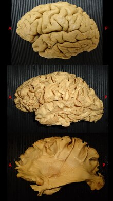
White matter dissection refers to a special anatomical technique able to reveal the subcortical organization of white matter fibers in the human or animal cadaver brain.
The first studies of cerebral white matter (WM) were described by Galen and by the subsequent efforts of Vesalius on human cadaver specimens.[2][3] The interest for the deep anatomy of the brain pushed anatomist during centuries to create and develop different techniques for specimen preparation and dissection in order to better reveal the complex white matter architectural organization.[2][3][4][5]
However, the biggest impact on the dissection of white matter anatomy was made by Joseph Klingler who developed a new method for specimens preparation and dissection. This technique became more feasible and widely used due to an increased quality of dissection and surprising quality of anatomical details.[4][5][6][7] Klingler developed a new method of brain fixation, by freezing already formalin-fixed brains before dissection.[6][7] First, the water crystallization induced by freezing disrupts the structure of the grey matter (which has a high water content). This process made possible to peel off the cortex from the brain surface without damaging the subcortical white matter organization underneath. Second, the freezing process along the WM fibers, induced a clear separation between them facilitating the dissection by progressive peeling of the fibers.[4][8][9]
White matter fibre dissection is nowadays considered as a valuable tool to enhance our knowledge about brain connectivity,[5][9][10][1] and has been used to validate tractographic results and vice versa with good consistency between the two techniques,[11] but also for neurosurgical training and neuroanatomical teaching.
References edit
- ^ a b Latini, Francesco; Hjortberg, Mats; Aldskogius, Håkan; Ryttlefors, Mats (September 2015). "The use of a cerebral perfusion and immersion–fixation process for subsequent white matter dissection". Journal of Neuroscience Methods. 253: 161–169. doi:10.1016/j.jneumeth.2015.06.019. PMID 26149289.
- ^ a b Clarke E, O’Malley CD. 1968. The Human Brain and Spinal Cord, A Historical Study Illustrated By Writings from Antiquity To The Twentieth Century. Berkeley and Los Angeles: University of California Press.[page needed]
- ^ a b Clarke, Edwin; O'Malley, Charles Donald (1996). The Human Brain and Spinal Cord: A Historical Study Illustrated by Writings from Antiquity to the Twentieth Century. Norman Publishing. ISBN 978-0-930405-25-0.[page needed]
- ^ a b c Türe, Uğur; Yaşargil, M. Gazi; Friedman, Allan H.; Al-Mefty, Ossama (August 2000). "Fiber Dissection Technique: Lateral Aspect of the Brain". Neurosurgery. 47 (2): 417–427. doi:10.1097/00006123-200008000-00028. PMID 10942015.
- ^ a b c Agrawal, Abhishek; Kapfhammer, Josef P; Kress, Annetrudi; Wichers, Hermann; Deep, Aman; Feindel, William; Sonntag, Volker K H; Spetzler, Robert F; Preul, Mark C (August 2011). "Josef Klinglerʼs Models of White Matter Tracts: Influences on Neuroanatomy, Neurosurgery, and Neuroimaging". Neurosurgery. 69 (2): 238–254. doi:10.1227/NEU.0b013e318214ab79. PMID 21368687.
- ^ a b Klingler, J (1935). "Erleichterung der makroskopischen Praeparation des Gehirns durch den Gefrierprozess" [Facilitation of the macroscopic preparation of the brain through the freezing process]. Schweizer Archiv für Neurologie und Psychiatrie (in German). 36: 247–256.
- ^ a b Ludwig E, Klinger J. 1956. Atlas Cerebri Humani. Basel: S. Karger.[page needed]
- ^ Fernández-Miranda, Juan C; Rhoton, Albert L; Alvarez-Linera, Juan; Kakizawa, Yukinari; Choi, Chanyoung; de Oliveira, Evandro P (June 2008). "Three-dimensional microsurgical and tractographic anatomy of the white matter of the human brain". Neurosurgery. 62 (6 Suppl 3): 989–1026, discussion 1026-8. doi:10.1227/01.neu.0000333767.05328.49. PMID 18695585.
- ^ a b Zemmoura, Ilyess; Blanchard, Emmanuelle; Raynal, Pierre-Ivan; Rousselot-Denis, Cécilia; Destrieux, Christophe; Velut, Stéphane (24 April 2015). "How Klingler's dissection permits exploration of brain structural connectivity? An electron microscopy study of human white matter". Brain Structure and Function. 221 (5): 2477–2486. doi:10.1007/s00429-015-1050-7. PMID 25905864.
- ^ Arnts, Hisse; Kleinnijenhuis, Michiel; Kooloos, Jan G.M.; Schepens-Franke, Annelieke N.; van Cappellen van Walsum, Anne-Marie (January 2014). "Combining fiber dissection, plastination, and tractography for neuroanatomical education: Revealing the cerebellar nuclei and their white matter connections". Anatomical Sciences Education. 7 (1): 47–55. doi:10.1002/ase.1385. PMID 23839938.
- ^ Zemmoura, Ilyess; Serres, Barthélémy; Andersson, Frédéric; Barantin, Laurent; Tauber, Clovis; Filipiak, Isabelle; Cottier, Jean-Philippe; Venturini, Gilles; Destrieux, Christophe (December 2014). "FIBRASCAN: A novel method for 3D white matter tract reconstruction in MR space from cadaveric dissection". NeuroImage. 103: 106–118. doi:10.1016/j.neuroimage.2014.09.016. PMID 25234114.