


Summary
The zygomatic nerve is a branch of the maxillary nerve (itself a branch of the trigeminal nerve (CN V)). It arises in the pterygopalatine fossa and enters the orbit through the inferior orbital fissure before dividing into its two terminal branches: the zygomaticotemporal nerve and zygomaticofacial nerve.
| Zygomatic nerve | |
|---|---|
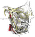
 Lateral view of the nerves of the orbit. The zygomatic nerve is visible at bottom centre branching from the maxillary nerve. | |
| Details | |
| From | maxillary nerve |
| To | zygomaticotemporal nerve communicating branch to lacrimal nerve |
| Innervates | skin over temporal bone and zygomatic bone |
| Identifiers | |
| Latin | nervus zygomaticus |
| TA98 | A14.2.01.056 |
| TA2 | 6231 |
| FMA | 52967 |
| Anatomical terms of neuroanatomy [edit on Wikidata] | |
Through its branches, the zygomatic nerve provides sensory invervation to skin over the zygomatic bone and the temporal bone. It also carries post-ganglionic parasympathetic axons to the lacrimal gland.
It may be blocked by anaesthetising the maxillary nerve.
Structure edit
Origin edit
The zygomatic nerve is a branch of the maxillary nerve (CN V2).[1][2] It arises at the pterygopalatine ganglion.[1]
Course edit
It exits from the pterygopalatine fossa through the inferior orbital fissure to enter the orbit.[1][3] In the orbit, it travels anteriorly along its lateral wall.[3]
Branches edit
Soon after the zygomatic nerve enters the orbit, it divides into its branches. These include:
- Zygomaticotemporal nerve[1]
- Zygomaticofacial nerve[1]
- A communicating branch to lacrimal nerve[1]
Variation edit
Sometimes, the zygomatic nerve does not branch within the orbit. Instead, it enters a single foramen in the zygomatic bone called the zygomatico-orbital foramen. In this case, it divides within the bone into the zygomaticotemporal nerve and the zygomaticofacial nerve.[4]
Function edit
The terminal branches of the zygomatic nerve contain sensory axons.[1] These provide sensation to the skin over the temporal bone and the zygomatic bone.[4]
The zygomatic nerve also carries postganglionic parasympathetic axons.[1] These axons have their cell bodies in the pterygopalatine ganglion. They travel from the ganglion to the zygomatic nerve, and then to the lacrimal nerve through a communicating branch. From the lacrimal nerve, they enter the lacrimal gland and provide secretomotor supply.[5]
Clinical significance edit
The zygomatic nerve can be blocked indirectly by anaesthetising the maxillary nerve (CN V2).[2] The zygomatic nerve and its branches may be damaged by a fracture to the zygomatic bone.[6]
Additional images edit
-
 The nerves of the scalp, face, and side of neck.
 -
 Branches of the trigeminal nerve. The zygomatic nerve is visible branching from the maxillary nerve and entering the orbit.


References edit
- ^ a b c d e f g h Rea, Paul (2016). "2 - Head". Essential Clinically Applied Anatomy of the Peripheral Nervous System in the Head and Neck. Academic Press. pp. 21–130. doi:10.1016/B978-0-12-803633-4.00002-8. ISBN 978-0-12-803633-4.
- ^ a b Pai, Umeshraya T.; Nayak, Rajeshri; Molloy, Robert E. (2005). "72 - Head and Neck Blocks". Essentials of Pain Medicine and Regional Anesthesia (2nd ed.). Churchill Livingstone. pp. 598–606. doi:10.1016/B978-0-443-06651-1.50076-9. ISBN 978-0-443-06651-1.
- ^ a b Forrester, John V.; Dick, Andrew D.; McMenamin, Paul G.; Roberts, Fiona; Pearlman, Eric (2016). "1 - Anatomy of the eye and orbit". The Eye - Basic Sciences in Practice (4th ed.). Saunders. pp. 1–102. doi:10.1016/B978-0-7020-5554-6.00001-0. ISBN 978-0-7020-5554-6.
- ^ a b Standring, Susan, ed. (2016). Gray's anatomy : the anatomical basis of clinical practice (41 ed.). Philadelphia: Elsevier. ISBN 978-0-7020-5230-9. OCLC 920806541.
- ^ Anderson, B. C.; McLoon, L. K. (2010). "Cranial Nerves and Autonomic Innervation in the Orbit". Encyclopedia of the Eye. Academic Press. pp. 537–548. doi:10.1016/B978-0-12-374203-2.00285-2. ISBN 978-0-12-374203-2.
- ^ Gellrich, Nils-Claudius Bernhard; Zimmerer, Rüdiger M. (2017). "7 - Surgical Management of Maxillary and Zygomatic Fractures". Maxillofacial Surgery. Vol. 1 (3rd ed.). Churchill Livingstone. pp. 93–132. doi:10.1016/B978-0-7020-6056-4.00007-1. ISBN 978-0-7020-6056-4.