
KNOWPIA
WELCOME TO KNOWPIA

Pterygoid canal 
Summary
The pterygoid canal (also vidian canal) is a passage in the sphenoid bone of the skull leading from just anterior to the foramen lacerum in the middle cranial fossa to the pterygopalatine fossa.[1]
| Pterygoid canal | |
|---|---|
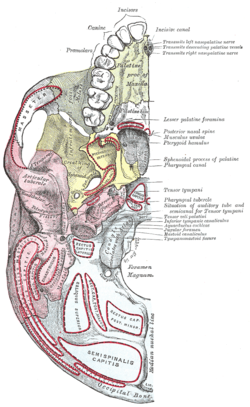 Base of skull. Inferior surface. (Sphenoid is yellow.) | |
 Sphenoid bone. Anterior and inferior surfaces. (Pterygoid c. labeled at center left.) | |
| Details | |
| Artery | artery of the pterygoid canal |
| Nerve | nerve of pterygoid canal |
| Identifiers | |
| Latin | canalis pterygoideus |
| TA98 | A02.1.05.053 |
| TA2 | 639 |
| FMA | 54756 |
| Anatomical terminology [edit on Wikidata] | |
It transmits the nerve of pterygoid canal (Vidian nerve), the artery of the pterygoid canal (Vidian artery), and the vein of the pterygoid canal (Vidian vein).
Structure edit
The pterygoid canal runs through the medial pterygoid plate of the sphenoid bone to the back wall of the pterygopalatine fossa.
Additional images edit
-
 Medial wall of left orbit.

External links edit
- Anatomy figure: 22:4b-08 at Human Anatomy Online, SUNY Downstate Medical Center
- cranialnerves at The Anatomy Lesson by Wesley Norman (Georgetown University) (VII)
References edit
- ^ Omami, Galal; Hewaidi, Gebril; Mathew, Reji (1 October 2011). "The neglected anatomical and clinical aspects of pterygoid canal: CT scan study". Surgical and Radiologic Anatomy. 33 (8): 697–702. doi:10.1007/s00276-011-0808-8. ISSN 1279-8517. PMID 21445687. S2CID 206948796. Retrieved 5 November 2022.