


-
 Phylogenetic reconstruction of the opsins. The outgroup contains other G protein-coupled receptors. The frame highlights the tetraopsins, which are expanded in the next image.
 -
 Phylogenetic reconstruction of the tetraopsins. The outgroup contains other G protein-coupled receptors including the other opsins. The frame highlights the chromopsins, which are expanded in the next image.
 -
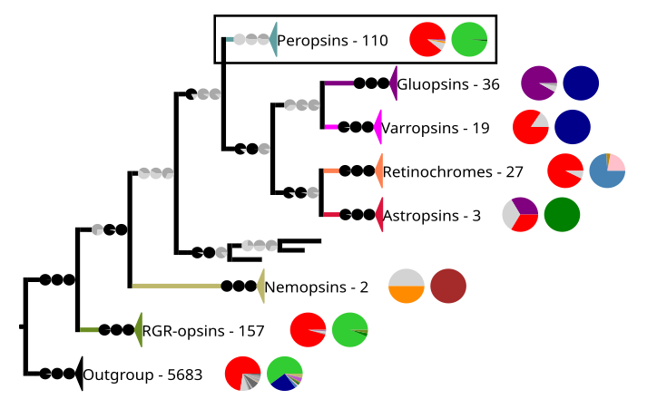 Phylogenetic reconstruction of the chromopsins. The outgroup contains other G protein-coupled receptors including the other opsins. The frame highlights the peropsins.

KNOWPIA
WELCOME TO KNOWPIA
RRH 
Summary
Peropsin, a visual pigment-like receptor, is a protein that in humans is encoded by the RRH gene.[5][6] It belongs like other animal opsins to the G protein-coupled receptors.[6] Even so, the first peropsins were already discovered in mice and humans in 1997,[5] not much is known about them.[7]
| RRH | |||||||||||||||||||||||||||||||||||||||||||||||||||
|---|---|---|---|---|---|---|---|---|---|---|---|---|---|---|---|---|---|---|---|---|---|---|---|---|---|---|---|---|---|---|---|---|---|---|---|---|---|---|---|---|---|---|---|---|---|---|---|---|---|---|---|
| Identifiers | |||||||||||||||||||||||||||||||||||||||||||||||||||
| Aliases | RRH, retinal pigment epithelium-derived rhodopsin homolog | ||||||||||||||||||||||||||||||||||||||||||||||||||
| External IDs | OMIM: 605224 MGI: 1097709 HomoloGene: 55977 GeneCards: RRH | ||||||||||||||||||||||||||||||||||||||||||||||||||
| |||||||||||||||||||||||||||||||||||||||||||||||||||
| |||||||||||||||||||||||||||||||||||||||||||||||||||
| |||||||||||||||||||||||||||||||||||||||||||||||||||
| |||||||||||||||||||||||||||||||||||||||||||||||||||
| |||||||||||||||||||||||||||||||||||||||||||||||||||
| Wikidata | |||||||||||||||||||||||||||||||||||||||||||||||||||
| |||||||||||||||||||||||||||||||||||||||||||||||||||
Photochemistry edit
Like most opsins, peropsins have in its seventh transmembrane domain a lysine corresponding to amino acid position 296 in cattle rhodopsin,[5][7] which is important for retinal binding and light sensing.[8]
In amphioxus, a cephalochordate, a peropsin binds in the dark-state all-trans-retinal instead of 11-cis-retinal,[9] as it is in cattle rhodopsin.[10][11][12][13][14] Therefore, peropsins have been suggested to be photoisomerases.[9]
Tissue localization edit
In mice, a peropsin is localized to the apical microvilli of the retinal pigment epithelium (RPE).[5] There, it regulates storage or the movement of vitamin A from the retina to the RPE.[15] A peropsin is also expressed in keratinocytes of the human skin. In keratinocyte cell culture, it reacts to UV light if retinal is supplied.[16] In chicken, a peropsin is expressed with an RGR-opsin in the pineal gland and the retina.[17]
Gene localization and structure edit
The human peropsin gene lies on chromosome 4 band 4q25 and has six introns[6][18] like RGR-opsins. However only two of these introns are inserted at the same place, which still indicates that peropsins and RGR-opsins are more closely related to each other than to the ciliary and rhabdomeric opsins.[18] This shared gene structure is also reflected in opsin phylogenies, where peropsins and RGR-opsins are in the same group: The chromopsins.[18][7][19][20]
Phylogeny edit
The peropsins are restricted to the craniates and the cephalochordates.[7] The craniates are the taxon that contains mammals and with them humans. The peropsins are one of the seven subgroups of the chromopsins. The other groups are the RGR-opsins, the retinochromes, the nemopsins, the astropsins, the varropsins, and the gluopsins.[7] The chromopsins are one of three subgroups of the tetraopsins (also known as RGR/Go or Group 4 opsins). The other groups are the neuropsins and the Go-opsins. The tetraopsins are one of the five major groups of the animal opsins, also known as type 2 opsins). The other groups are the ciliary opsins (c-opsins, cilopsins), the rhabdomeric opsins (r-opsins, rhabopsins), the xenopsins, and the nessopsins. Four of these subclades occur in Bilateria (all but the nessopsins).[7][19] However, the bilaterian clades constitute a paraphyletic taxon without the opsins from the cnidarians.[7][19][20][21]
The phylogenetic relationship of the peropsins to the other opsins


In the phylogeny above, Each clade contains sequences from opsins and other G protein-coupled receptors. The number of sequences and two pie charts are shown next to the clade. The first pie chart shows the percentage of a certain amino acid at the position in the sequences corresponding to position 296 in cattle rhodopsin. The amino acids are color-coded. The colors are red for lysine (K), purple for glutamic acid (E), orange for arginine (R), dark and mid-gray for other amino acids, and light gray for sequences that have no data at that position. The second pie chart gives the taxon composition for each clade, green stands for craniates, dark green for cephalochordates, mid green for echinoderms, brown for nematodes, pale pink for annelids, dark blue for arthropods, light blue for mollusks, and purple for cnidarians. The branches to the clades have pie charts, which give support values for the branches. The values are from right to left SH-aLRT/aBayes/UFBoot. The branches are considered supported when SH-aLRT ≥ 80%, aBayes ≥ 0.95, and UFBoot ≥ 95%. If a support value is above its threshold the pie chart is black otherwise gray.[7]
Clinical significance edit
Since RGR-opsin may be associated with retinitis pigmentosa,[22] which is like peropsin also expressed in the retinal pigment epithelium, peropsin was screened for a link with retinitis pigmentosa.[23] However, no link could be established.[23][24]
References edit
- ^ a b c GRCh38: Ensembl release 89: ENSG00000180245 – Ensembl, May 2017
- ^ a b c GRCm38: Ensembl release 89: ENSMUSG00000028012 – Ensembl, May 2017
- ^ "Human PubMed Reference:". National Center for Biotechnology Information, U.S. National Library of Medicine.
- ^ "Mouse PubMed Reference:". National Center for Biotechnology Information, U.S. National Library of Medicine.
- ^ a b c d Sun H, Gilbert DJ, Copeland NG, Jenkins NA, Nathans J (September 1997). "Peropsin, a novel visual pigment-like protein located in the apical microvilli of the retinal pigment epithelium". Proceedings of the National Academy of Sciences of the United States of America. 94 (18): 9893–9898. Bibcode:1997PNAS...94.9893S. doi:10.1073/pnas.94.18.9893. PMC 23288. PMID 9275222.
- ^ a b c "Entrez Gene: RRH retinal pigment epithelium-derived rhodopsin homolog".
- ^ a b c d e f g h Gühmann M, Porter ML, Bok MJ (August 2022). "The Gluopsins: Opsins without the Retinal Binding Lysine". Cells. 11 (15): 2441. doi:10.3390/cells11152441. PMC 9368030. PMID 35954284.
 Material was copied and adapted from this source, which is available under a Creative Commons Attribution 4.0 International License.
- ^ Leung NY, Thakur DP, Gurav AS, Kim SH, Di Pizio A, Niv MY, et al. (April 2020). "Functions of Opsins in Drosophila Taste". Current Biology. 30 (8): 1367–1379.e6. Bibcode:2020CBio...30E1367L. doi:10.1016/j.cub.2020.01.068. PMC 7252503. PMID 32243853.
- ^ a b Koyanagi M, Terakita A, Kubokawa K, Shichida Y (November 2002). "Amphioxus homologs of Go-coupled rhodopsin and peropsin having 11-cis- and all-trans-retinals as their chromophores". FEBS Letters. 531 (3): 525–528. doi:10.1016/s0014-5793(02)03616-5. PMID 12435605. S2CID 11669142.
- ^ Wald G (July 1934). "Carotenoids and the Vitamin A Cycle in Vision". Nature. 134 (3376): 65. Bibcode:1934Natur.134...65W. doi:10.1038/134065a0. S2CID 4022911.
- ^ Wald G, Brown PK, Hubbard R, Oroshnik W (July 1955). "Hindered Cis Isomers of Vitamin A and Retinene: The Structure of the Neo-B Isomer". Proceedings of the National Academy of Sciences of the United States of America. 41 (7): 438–451. Bibcode:1955PNAS...41..438W. doi:10.1073/pnas.41.7.438. PMC 528115. PMID 16589696.
- ^ Brown PK, Wald G (October 1956). "The neo-b isomer of vitamin A and retinene". The Journal of Biological Chemistry. 222 (2): 865–877. doi:10.1016/S0021-9258(20)89944-X. PMID 13367054.
- ^ Oroshnik W (June 1956). "The Synthesis and Configuration of Neo-B Vitamin A and Neoretinine b". Journal of the American Chemical Society. 78 (11): 2651–2652. doi:10.1021/ja01592a095.
- ^ Oroshnik W, Brown PK, Hubbard R, Wald G (September 1956). "Hindered Cis Isomers of Vitamin A and Retinene: The Structure of the Neo-B Isomer". Proceedings of the National Academy of Sciences of the United States of America. 42 (9): 578–580. Bibcode:1956PNAS...42..578O. doi:10.1073/pnas.42.9.578. PMC 534254. PMID 16589909.
- ^ Cook JD, Ng SY, Lloyd M, Eddington S, Sun H, Nathans J, et al. (December 2017). "Peropsin modulates transit of vitamin A from retina to retinal pigment epithelium". The Journal of Biological Chemistry. 292 (52): 21407–21416. doi:10.1074/jbc.M117.812701. PMC 5766940. PMID 29109151.
- ^ Toh PP, Bigliardi-Qi M, Yap AM, Sriram G, Stelmashenko O, Bigliardi P (December 2016). "Expression of peropsin in human skin is related to phototransduction of violet light in keratinocytes". Experimental Dermatology. 25 (12): 1002–1005. doi:10.1111/exd.13226. PMID 27676658. S2CID 1373924.
- ^ Bailey MJ, Cassone VM (March 2004). "Opsin photoisomerases in the chick retina and pineal gland: characterization, localization, and circadian regulation". Investigative Ophthalmology & Visual Science. 45 (3): 769–775. doi:10.1167/iovs.03-1125. PMID 14985289.
- ^ a b c Bellingham J, Wells DJ, Foster RG (January 2003). "In silico characterisation and chromosomal localisation of human RRH (peropsin)--implications for opsin evolution". BMC Genomics. 4 (1): 3. doi:10.1186/1471-2164-4-3. PMC 149353. PMID 12542842.
- ^ a b c Ramirez MD, Pairett AN, Pankey MS, Serb JM, Speiser DI, Swafford AJ, et al. (26 October 2016). "The last common ancestor of most bilaterian animals possessed at least 9 opsins". Genome Biology and Evolution: evw248. doi:10.1093/gbe/evw248. PMC 5521729. PMID 27797948.
- ^ a b Porter ML, Blasic JR, Bok MJ, Cameron EG, Pringle T, Cronin TW, et al. (January 2012). "Shedding new light on opsin evolution". Proceedings. Biological Sciences. 279 (1726): 3–14. doi:10.1098/rspb.2011.1819. PMC 3223661. PMID 22012981.
- ^ Liegertová M, Pergner J, Kozmiková I, Fabian P, Pombinho AR, Strnad H, et al. (July 2015). "Cubozoan genome illuminates functional diversification of opsins and photoreceptor evolution". Scientific Reports. 5: 11885. Bibcode:2015NatSR...511885L. doi:10.1038/srep11885. PMC 5155618. PMID 26154478.
- ^ Morimura H, Saindelle-Ribeaudeau F, Berson EL, Dryja TP (December 1999). "Mutations in RGR, encoding a light-sensitive opsin homologue, in patients with retinitis pigmentosa". Nature Genetics. 23 (4): 393–394. doi:10.1038/70496. PMID 10581022. S2CID 35176366.
- ^ a b Ksantini M, Sénéchal A, Humbert G, Arnaud B, Hamel CP (March 2007). "RRH, encoding the RPE-expressed opsin-like peropsin, is not mutated in retinitis pigmentosa and allied diseases" (PDF). Ophthalmic Genetics. 28 (1): 31–37. doi:10.1080/13816810701202052. PMID 17454745. S2CID 225451.
- ^ Rivolta C, Berson EL, Dryja TP (December 2006). "Mutation screening of the peropsin gene, a retinal pigment epithelium specific rhodopsin homolog, in patients with retinitis pigmentosa and allied diseases". Molecular Vision. 12: 1511–1515. PMID 17167409.
This article incorporates text from the United States National Library of Medicine, which is in the public domain.